In the white matter of the frontal and parietal lobes. White matter of the brain: structure, functions
White matter hemispheres of the brain consists of three types of fibers - association, connecting separate areas the cortex of the cerebral hemispheres within just one hemisphere, commissural - connecting the cerebral hemispheres with each other and projection - conducting pathways of analyzers that carry out bilateral communication of the cerebral cortex with the underlying formations.
Internal capsule and semioval center. The internal capsule is a compact cluster of pathways going to the cortex and from the cortex to the underlying parts of the central nervous system. From the outside it borders on the lenticular nucleus, and from the inside on the optic tubercle and the caudate body.
The pathways are located in the internal capsule in a certain order. In its anterior thigh there are pathways connecting the frontal lobe of the brain with the cerebellum and with the optic thalamus. In the knee of the internal capsule there are corticonuclear pathways to the nuclei of the motor cranial nerves. The most anterior sections of this segment are occupied by fibers for combined eye movements.
In the posterior thigh of the internal capsule, the pathways lie in the following order. Its anterior sections are occupied by the pyramidal fasciculus. In the segment of the pyramidal tract passing through the internal capsule, the fibers are located in such a way that in front, directly adjacent to the corticobulbar tracts, lie the pyramidal fibers for the neck and arm, and more posteriorly for the trunk and legs. In both the foot and hand bundles, the finger conductors are behind the others, but the boundary between groups of conductors is usually blurred and the fibers are partially mixed. Here, in addition to the pyramidal fibers, there are cortico-rubral and thalapallidal connections. This should be kept in mind because any pathological focus in this place, in addition to the pyramidal ones, usually affects these connections. Posterior to the pyramidal conductors are sensory pathways running from the thalamus optic to the cerebral cortex.
Further, posteriorly, the visual pathways are located and, finally, in the pars sublenticularis there are the auditory pathway and the pathway connecting the temporal and occipital regions of the cortex with the cerebellum through the pons. As can be seen from the above, in the internal capsule the pathways are located in a certain sequence: connections of the frontal lobe with the underlying formations are located more orally, connections of the parietal lobe with the underlying formations are located posterior to them, and, finally, the caudal parts of the capsule are occupied by connections of the occipital and temporal lobes with the underlying formations. formations. Knowledge of the topography of the conductive pathways located in the capsule is necessary for topical diagnosis of its lesions. In this case, we must keep in mind the following circumstance. In the inner capsule, all conductors lie compactly on a fairly limited space, as a result of which a pathological focus in the internal capsule (for example, hemorrhage) simultaneously affects a number of conducting systems. This explains the massiveness of symptoms in capsular localizations of the process.
A different picture is observed with damage to the white matter, located under the cerebral cortex, down to the level of the subcortical nodes and known as centrum semiovale and corona radiata. Here, the sensitive conductors on the way from the capsule to the cortex begin to fan out, and even more so the closer to the cortex. On the contrary, the pyramidal tracts along the path from the cortex to the capsule begin to converge in a fan-shaped manner, and the closer to the capsule, the more so. This creates conditions under which pathological lesions in the centrum semiovale, other things being equal, simultaneously affect fewer conductors and cause less massive syndromes than with capsular lesions. The most commonly affected areas are the posterior hip and knee of the internal capsule. When the knee of the internal capsule is damaged, the corticobulbar pathways that send motor impulses to the nuclei of the motor cranial nerves are affected. But since most of these nerves receive bilateral corticonuclear innervation, only those that are connected to one opposite hemisphere of the brain are affected. The patient will experience central paralysis of the XII and VII nerves on the side opposite to the lesion. With bilateral damage to the knee of the internal capsule, the patient develops pseudobulbar palsy. Isolated damage to the conductors located in the knee of the internal capsule is rare. In most cases, it is combined with damage to the pyramidal fasciculus, and often to other conductors located in the posterior thigh of the internal capsule. In these cases, the patient, in addition to disruption of the supranuclear innervation of the VII and XII nerves, experiences spastic (central) hemiplegia on the opposite side. Capsular hemiplegia is characterized by a more or less uniform distribution of paralysis in the arm and leg, as well as a peculiar posture of the affected limbs. With it, the arm is abducted from the body and bent at the elbow joint, the hand is drained and bent. The fingers are also bent. The leg is extended at the hip and knee joints and adducted. The foot is flexed and slightly supinated. When walking, the patient, due to the “lengthening” of the leg, abducts it, describing it in a semicircle. This position, due to the selective distribution of muscle hypertension, is called the Wernicke-Mann position.
In paralyzed limbs, the distal parts suffer more. Movements of the trunk with unilateral lesions of the capsule are not noticeably impaired due to the double pyramidal innervation of the muscles of the trunk. Synkinesis, or pathological conjugate movements, are observed in paralyzed limbs.
If the pathological process, in addition to the pyramidal tract, also involves sensitive conductors, then the patient’s paralyzed limbs also suffer from sensitivity. Sensory impairment is more or less evenly distributed over the entire half of the body, with the arm being slightly affected. more legs, distal sections are larger than proximal ones. Of all types of sensitivity, deep sensitivity is most affected. Typically, with capsular localizations of the process, sensory disorders are less constant and less persistent than motor disorders.
When the visual pathways lying behind the conductors of general sensitivity are involved in the process, the patient develops homonymous hemianopsia - the halves of the visual fields opposite to the lesion are lost. Most often, hemianopia in these cases occurs as a negative scotoma (the patient does not notice the visual defect). When the “blind” halves are illuminated, the reaction of the pupils is preserved.
The section of the internal capsule, where the central auditory tract is located, is rarely affected. Only more subtle methods can detect bilateral hearing loss, more on the side opposite to the lesion.
It should be borne in mind that with capsular localizations of the process, only symptoms of prolapse are observed; There are no symptoms of irritation (motor, sensory, visual, etc.). Damage to the centrum semiovale, like damage to the internal capsule, is accompanied by disturbances in movement, sensitivity, etc. due to damage to pathways running in a centrifugal and centripetal direction relative to the cortex. However, the clinical symptoms are distinguished by a certain originality and consist in the fact that the symptoms of damage to the semi-oval center contain both capsular and cortical features, depending on the degree of damage. Thus, when the center semiovale is affected, a combination of symptoms of prolapse and symptoms of irritation (motor or sensory) is often encountered. In contrast to capsular localizations, uneven hemiplegia is observed, often approaching the type of cortical monoplegia. Sensitivity disorders are of the same nature: the area of distribution of these disorders is smaller than with capsular disorders, sensitivity suffers much more in the arm (more often) or in the leg. Here there are conditions under which dissociation of motor, sensory and other disorders occurs more often than simultaneous damage to the motor, sensory and visual pathways, as well as supranuclear innervation of the cranial nerves, as with damage to the internal capsule. Damage to the parts of the centrum semiovale closest to the cortex can affect not only the projection pathways, but also the commissural and association fibers lying directly under the cortex. And then the clinical picture can be supplemented by symptoms of a violation of higher cortical functions (speech disorders, apraxia, etc.).
Commissural fibers. Commissural fibers, concentrated mainly in the corpus callosum, connect the frontal, parietal, temporal and occipital lobes of both hemispheres. Therefore, in lesion syndromes corpus callosum Depending on the location of its damage, symptoms of damage to these areas of the brain are included to varying degrees. Often, when the corpus callosum is damaged, apraxia is observed in the clinic, limited only to the left hand. This selectivity of apractical disorders is explained by the fact that when the corpus callosum is damaged, the connection between the left parietal region and the right hemisphere, associated with the motor functions of the left hand, is disrupted.
Association fibers. Damage to association fibers causes symptoms of dysfunction of the cerebral cortex.
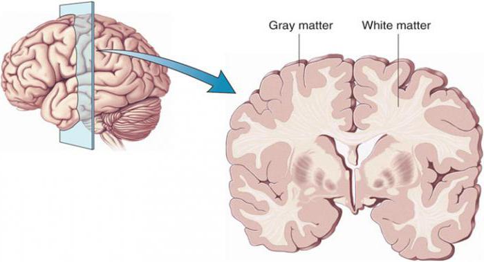
In the brain, gray and white matter are distinguished, but their distribution here is much more complex than in the spinal cord. Most of the gray matter of the brain is located on the surface big brain and the cerebellum, forming their cortex. The smaller part forms numerous subcortical nuclei, surrounded by white matter. All gray matter nuclei consist of multipolar neurons.
Gray matter (substantia grisea) contains the bodies of neurons, from which the nuclei of the central nervous system are formed. (nuclei) and bark (cortex). White matter (substantia alba) consists of processes of neurons that form bundles (fasciculi) and tracts (tractus), which are links in the pathways of the central nervous system.
The brain consists of gray and white matter. White matter occupies the entire space between the gray matter of the cerebral cortex and the basal ganglia. The surface of the hemisphere, the cloak (pallium), is formed by a uniform layer of gray matter 1.3 - 4.5 mm thick, containing nerve cells.
White matter has four parts:
1. central substance of the corpus callosum, internal capsule and long associative fibers;
2. radiant crown (corona radiata), formed by radiating fibers entering and leaving the internal capsule (capsula interna);
3. area of white matter in the outer parts of the hemisphere - semi-oval center (centrum semiovale);
4. white matter in the gyri between the sulci.
Nerve fibers of white matter are divided into projection, associative and commissural.
The white matter of the hemispheres is formed by nerve fibers connecting the cortex of one gyrus with the cortex of other gyri of its and the opposite hemispheres, as well as with underlying formations.
Two cerebral commissures, commissura anterior and commissura fornicis, are much smaller in size and belong to olfactory brain rhinencephalon and connect: commissura anterior - olfactory lobes and both parahippocampal gyri, commissura fornicis - hippocampi.
Commissural fibers, which are part of the cerebral commissures, or commissures, connect not only symmetrical points, but also the cortex belonging to different lobes of the opposite hemispheres.
Association fibers connect different parts of the cortex of the same hemisphere.
Associative fibers are divided into short and long.
Short fibers connect neighboring convolutions in the form of arcuate bundles.
Long association fibers connect areas that are more distant from each other
Projection fibers connect the cerebral cortex with the underlying formations, and through them with the periphery.
On a frontal section of the brain, the internal capsule looks like an oblique white stripe that continues into the cerebral peduncle.
In the internal capsule, the anterior leg (crus anterius) is distinguished - between the caudate nucleus and the anterior half of the inner surface of the lentiform nucleus, as well as the posterior leg (crus posterius) - between the thalamus and the posterior half of the lenticular nucleus and the knee (genu). Projection fibers according to their length can be divided into the following three systems:
Fibrae thalamocorticalis et corticothalamici - fibers from the thalamus to the cortex and back from the cortex to the thalamus; conducting excitation towards the cortex and centrifugal (descending, corticofugal, efferent).
Tractus corticonuclearis - pathways to the motor nuclei of the cranial nerves. Since all motor fibers are collected in a small space in the internal capsule (the knee and the anterior two-thirds of its posterior leg), if they are damaged in this place, unilateral paralysis of the opposite side of the body is observed.
Tractus corticospinalis (pyramidalis) conducts motor volitional impulses to the muscles of the trunk and limbs.
Tractus corticopontini - pathways from the cerebral cortex to the pontine nuclei. Using these pathways, the cerebral cortex has an inhibitory and regulatory effect on the activity of the cerebellum.
Projection fibers in the white matter of the hemisphere closer to the cortex form the corona radiata, and then main part they converge into the internal capsule, which is a layer of white matter between the lentiform nucleus (nucleus lentiformis), the caudate nucleus (nucleus caudatus) and the thalamus (thalamus).
Now let's look at the gray matter. The surface of the cloak has a very complex drawing, consisting of alternating various directions grooves and ridges between them, called gyri.
Deep, permanent grooves are used to divide each hemisphere into large areas called lobes; the latter, in turn, are divided into lobules and convolutions.
The size and shape of the grooves are subject to significant individual fluctuations, as a result of which not only the brain different people, but even the hemispheres of the same individual are not quite similar in the pattern of grooves.
There are five lobes of the hemisphere: frontal (lobus frontalis), parietal (lobus parietalis), temporal (lobus temporalis), occipital (lobus occipitalis) and a lobe hidden at the bottom lateral sulcus- the so-called islet (insula).
The central sulcus (sulcus cenrtalis) begins at the upper edge of the hemisphere and goes forward and down. The part of the hemisphere located in front of the central sulcus belongs to the frontal lobe. The part of the brain surface lying posterior to the central sulcus constitutes the parietal lobe. The posterior border of the parietal lobe is the end of the parieto-occipital sulcus (sulcus parietooccipitalis), located on medial surface hemispheres.
Frontal lobe. In the posterior part of the outer surface of this lobe the sulcus precentralis runs almost parallel to the direction of the sulcus centralis. Two grooves run from it in the longitudinal direction: sulcus frontalis superior et sulcus frontalis inferior. Due to this, the frontal lobe is divided into four convolutions.
The vertical gyrus, gyrus precentralis, is located between the central and precentral sulci Superolateral surface the hemisphere is delimited into lobes by three grooves: the lateral, central and upper end of the parieto-occipital groove.
The lateral sulcus (sulcus cerebri lateralis) begins on the basal surface of the hemisphere from the lateral fossa and then passes to the superolateral surface
The lobe consists of a number of convolutions, called in some places lobules, which are limited by the grooves of the brain surface.
The horizontal convolutions of the frontal lobe are: superior frontal (gyrus frontalis superior), middle frontal (gyrus frontalis medius) and inferior frontal (gyrus frontalis inferior).
Temporal lobe. The lateral surface of this lobe has three longitudinal convolutions, delimited from each other by sulcus temporalis superior and sulcus temporalis inferior. The gyrus temporalis medius extends between the superior and inferior temporal grooves. Below it runs the gyrus temporalis inferior.
Occipital lobe. The grooves on the lateral surface of this lobe are variable and inconsistent. Of these, the transversely running sulcus occipitalis transversus is distinguished, usually connecting to the end of the interparietal sulcus.
Parietal lobe. On it, approximately parallel to the central groove, there is a sulcus postcentralis, usually merging with the sulcus intraparietalis, which runs in a horizontal direction. Depending on the location of these grooves, the parietal lobe is divided into three gyri.
The vertical gyrus, gyrus postcentralis, runs behind the central sulcus in the same direction as the precentral gyrus. Above the interparietal sulcus is the superior parietal gyrus, or lobule (lobulus parietalis superior), below - lobulus parietalis inferior.
Island. This lobe has the shape of a triangle. The surface of the insula is covered with short convolutions.
The lower surface of the hemisphere in that part that lies in front of the lateral fossa belongs to the frontal lobe.
On the posterior portion of the basal surface of the hemisphere, two grooves are visible: the sulcus occipitotemporalis, running in the direction from the occipital pole to the temporal and limiting the gyrus occipitotemporalis lateralis, and the sulcus collateralis running parallel to it. Here the sulcus olfactorius runs parallel to the medial edge of the hemisphere. Parallel to and above this groove, the sulcus cinguli runs along the medial surface of the hemisphere. Between them is the gyrus occipitotemporalis medialis.
There are two gyri located medially from the collateral sulcus: between the posterior section of this sulcus and the sulcus calcarinus lies the gyrus lingualis; between the anterior section of this groove and the deep sulcus hippocampi lies the gyrus parahippocampalis.
The gyrus adjacent to the brain stem is already located on the medial surface of the hemisphere.
Behind the precuneus lies a separate area of the cortex related to occipital lobe, - wedge (cuneus). Between the lingual sulcus and the sulcus of the corpus callosum stretches the cingulate gyrus (gyrus cinguli), which, through the isthmus (isthmus), continues into the parahippocampal gyrus, ending with the hook (uncus). Gyrus cinguli, isthmus and gyrus parahippocampalis form together a vaulted gyrus (gyrus fornicatus), which describes an almost complete circle, open only below and in front.
On the medial surface of the hemisphere there is a groove of the corpus callosum (sulcus corpori callosi), running directly above the corpus callosum and continuing with its posterior end into the deep sulcus hippocampi, which is directed forward and downward.
The paracentral lobule (lobulus paracentralis) is a small area above the lingual sulcus. From the paracentral lobule there is a quadrangular surface (the so-called precuneus, precuneus). It belongs to the parietal lobe. The vaulted gyrus is not related to any of the cloak lobes. It belongs to the limbic region. The limbic region is part of the neocortex of the cerebral hemispheres, occupying the cingulate and parahippocampal gyri; part of the limbic system. Pulling apart the edge of the sulcus hippocampi, one can see a narrow jagged gray stripe, which is a rudimentary gyrus of the gyrus dentatus.
Abstracts The brain consists of gray and white matter. White matter occupies the entire space between the gray matter of the cerebral cortex and the basal ganglia. The surface of the hemisphere, the cloak (pallium), is formed by a uniform layer of gray matter 1.3 - 4.5 mm thick, containing nerve cells. First, let's look at white matter. White matter has four parts: 1) the central substance of the corpus callosum, internal capsule and long associative fibers. 2) radiant crown (corona radiata), formed by radiating fibers entering and leaving the internal capsule (capsula interna); 3) the area of white matter in the outer parts of the hemisphere - the semi-oval center (centrum semiovale); 4) white matter in the gyri between the sulci; Nerve fibers of white matter are divided into projection, associative and commissural. The white matter of the hemispheres is formed by nerve fibers connecting the cortex of one gyrus with the cortex of other gyri of its and the opposite hemispheres, as well as with underlying formations. Two brain commissures, commissura anterior and commissura fornicis, are much smaller in size and belong to the olfactory brain rhinencephalon and connect: commissura anterior - olfactory lobes and both parahippocampal gyri, commissura fornicis - hippocampi. Most of the commissural fibers are part of the corpus callosum, which connects the parts of both hemispheres belonging to the brain. Commissural fibers, which are part of the cerebral commissures, or commissures, connect not only symmetrical points, but also the cortex belonging to different lobes of the opposite hemispheres. Association fibers connect different parts of the cortex of the same hemisphere. Associative fibers are divided into short and long. Short fibers connect neighboring convolutions in the form of arcuate bundles. Long association fibers connect areas of the cortex that are more distant from each other. Projection fibers connect the cerebral cortex with the underlying formations, and through them with the periphery. These fibers are divided into centripetal (ascending, corticopetal, afferent). On a frontal section of the brain, the internal capsule looks like an oblique white stripe that continues into the cerebral peduncle. In the internal capsule, the anterior leg (crus anterius) is distinguished, between the caudate nucleus and the anterior half of the inner surface of the lentiform nucleus, the posterior leg (crus posterius), between the thalamus and the posterior half of the lentiform nucleus and the knee (genu), lying at the inflection point between both parts of the inner capsule. Projection fibers can be divided according to their length into the following three systems, starting with the longest: 1. Fibrae thalamocorticalis et corticothalamici - fibers from the thalamus to the cortex and back from the cortex to the thalamus. Conducting excitation towards the cortex, and centrifugal (descending, corticofugal, efferent). 2. Tractus corticonuclearis - pathways to the motor nuclei of the cranial nerves. Since all motor fibers are collected in a small space in the internal capsule (the knee and the anterior two-thirds of its posterior leg), if they are damaged in this place, unilateral paralysis of the opposite side of the body is observed. 3. Tractus corticospinalis (pyramidalis) conducts motor volitional impulses to the muscles of the trunk and limbs. 4. Tractus corticopontini - paths from the cerebral cortex to the pontine nuclei. Using these pathways, the cerebral cortex has an inhibitory and regulatory effect on the activity of the cerebellum. Projection fibers in the white matter of the hemisphere closer to the cortex form the corona radiata, and then the main part of them converges into the internal capsule, which is a layer of white matter between the lentiform nucleus (nucleus lentiformis) on one side, and the caudate nucleus (nucleus caudatus) and thalamus ( thalamus) - on the other. Now let's look at the gray matter. The surface of the cloak has a very complex pattern, consisting of furrows alternating in different directions and ridges between them, called convolutions, gyri. Deep permanent grooves are used to divide each hemisphere into large areas called lobes, lobi; the latter, in turn, are divided into lobules and convolutions. The size and shape of the grooves are subject to significant individual fluctuations, as a result of which not only the brains of different people, but even the hemispheres of the same individual are not quite similar in the pattern of the grooves. There are five lobes of the hemisphere: frontal (lobus frontalis), parietal (lobus parietalis), temporal (lobus temporalis), occipital (lobus occipitalis) and a lobe hidden at the bottom of the lateral sulcus, the so-called insula. The central sulcus (sulcus cenrtalis) begins at the upper edge of the hemisphere and goes forward and down. The area of the hemisphere located in front of the central sulcus. Refers to the frontal lobe. The part of the brain surface lying posterior to the central sulcus constitutes the parietal lobe. The posterior border of the parietal lobe is the end of the parieto-occipital sulcus (sulcus parietooccipitalis), located on the medial surface of the hemisphere. Frontal lobe. In the posterior part of the outer surface of this lobe the sulcus precentralis runs almost parallel to the direction of the sulcus centralis. Two grooves run from it in the longitudinal direction: sulcus frontalis superior et sulcus frontalis inferior. Due to this, the frontal lobe is divided into four convolutions. The vertical gyrus, gyrus precentralis, is located between the central and precentral sulci. The superior lateral surface of the hemisphere is delimited into lobes by three sulci: the lateral, central and the upper end of the parieto-occipital sulcus. The lateral sulcus (sulcus cerebri lateralis) begins on the basal surface of the hemisphere from the lateral fossa and then passes to the superolateral surface The lobe consists of a number of convolutions, called in some places lobules, which are limited by the grooves of the brain surface. The horizontal convolutions of the frontal lobe are: superior frontal (gyrus frontalis superior), middle frontal (gyrus frontalis medius) and inferior frontal (gyrus frontalis inferior). Temporal lobe. The lateral surface of this lobe has three longitudinal convolutions, delimited from each other by sulcus temporalis superior and sulcus temporalis inferior. The gyrus temporalis medius extends between the superior and inferior temporal grooves. Below it runs the gyrus temporalis inferior. Occipital lobe. The grooves on the lateral surface of this lobe are variable and inconsistent. Of these, the transversely running sulcus occipitalis transversus is distinguished, usually connecting to the end of the interparietal sulcus. Parietal lobe. On it, approximately parallel to the central groove, there is a sulcus postcentralis, usually merging with the sulcus intraparietalis, which runs in a horizontal direction. Depending on the location of these grooves, the parietal lobe is divided into three gyri. The vertical gyrus, gyrus postcentralis, runs behind the central sulcus in the same direction as the precentral gyrus. Above the interparietal sulcus is the superior parietal gyrus, or lobule (lobulus parietalis superior), below - lobulus parietalis inferior. Island. This lobe has the shape of a triangle. The surface of the insula is covered with short convolutions. The lower surface of the hemisphere in that part that lies anterior to the lateral fossa belongs to the frontal lobe. On the posterior portion of the basal surface of the hemisphere, two grooves are visible: the sulcus occipitotemporalis, running in the direction from the occipital pole to the temporal and limiting the gyrus occipitotemporalis lateralis, and the sulcus collateralis running parallel to it. Here the sulcus olfactorius runs parallel to the medial edge of the hemisphere. Parallel to and above this groove, the sulcus cinguli runs along the medial surface of the hemisphere. Between them is the gyrus occipitotemporalis medialis. There are two gyri located medially from the collateral sulcus: between the posterior section of this sulcus and the sulcus calcarinus lies the gyrus lingualis; between the anterior section of this groove and the deep sulcus hippocampi lies the gyrus parahippocampalis. The gyrus adjacent to the brain stem is already located on the medial surface of the hemisphere. Behind the precuneus lies a separate area of the cortex belonging to the occipital lobe - the cuneus. Between the lingual sulcus and the sulcus of the corpus callosum stretches the cingulate gyrus (gyrus cinguli), which, through the isthmus (isthmus), continues into the parahippocampal gyrus, ending with the hook (uncus). Gyrus cinguli, isthmus and gyrus parahippocampalis form together a vaulted gyrus (gyrus fornicatus), which describes an almost complete circle, open only below and in front. On the medial surface of the hemisphere there is a groove of the corpus callosum (sulcus corpori callosi), running directly above the corpus callosum and continuing with its posterior end into the deep sulcus hippocampi, which is directed forward and downward. The paracentral lobule (lobulus paracentralis) is a small area above the lingual sulcus. From the paracentral lobule there is a quadrangular surface (the so-called precuneus, precuneus). It belongs to the parietal lobe. The vaulted gyrus is not related to any of the cloak lobes. It belongs to the limbic region. The limbic region is part of the neocortex of the cerebral hemispheres, occupying the cingulate and parahippocampal gyri; part of the limbic system. Pulling apart the edge of the sulcus hippocampi, one can see a narrow jagged gray stripe, which is a rudimentary gyrus of the gyrus dentatus. Bibliography 1. M.G. Prives, N.K. Lysenkov, V.I. Bushkovich. Human anatomy. M., 1985 2. Great medical encyclopedia. vol. 11, M., 1979 3. Great medical encyclopedia. vol. 6, M., 1977Gray and white matter of the brain
The human brain consists of white and gray matter. The first is everything that is filled between the gray matter on the cortex and on the surface there is a uniform layer of gray matter with nerve cells, the thickness of which is up to four and a half millimeters.
Let's study in more detail what gray and white matter is in the brain.
What are these substances made of?
The substance of the central nervous system is of two types: white and gray.
White matter consists of many nerve fibers and nerve cell processes, the membrane of which is white.
Gray matter consists of processes. Nerve fibers connect different parts of the central nervous system and nerve centers.
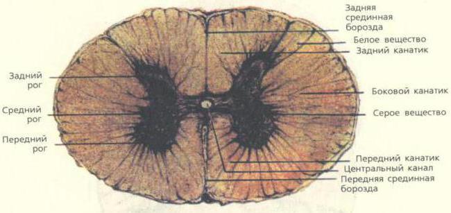
Gray and white matter of the spinal cord
The heterogeneous substance of this organ is gray and white. The first is formed by a huge number of neurons, which are concentrated in nuclei and come in three types:
- radicular cells;
- tufted neurons;
- internal cells.
The white matter of the spinal cord surrounds the gray matter. It includes nerve processes that make up three fiber systems:
- intercalary and afferent neurons connecting different parts of the spinal cord;
- sensory afferents, which are long centripetal;
- motor afferent or long centrifugal.
Medulla
From the anatomy course we know that the spinal cord passes into the medulla oblongata. The part of this brain at the top is thicker than at the bottom. Its average length is 25 millimeters, and its shape resembles a truncated cone.
It develops gravitational and auditory organs associated with breathing and blood circulation. Therefore, the nuclei of gray matter here regulate balance, metabolism, blood circulation, breathing, and coordination of movements.
hindbrain
This brain consists of the pons and the cerebellum. Let's look at the gray and white matter in them. The bridge is a large white ridge on the back side of the base. On the one hand, its border with the cerebral peduncles is pronounced, and on the other, with the medulla oblongata. If you make a cross section, the white matter of the brain and the gray nucleus will be visible very clearly. Transverse fibers divide the bridge into ventral and dorsal sections. In the ventral part, the white matter of the pathways is mainly present, and the gray matter forms its nuclei here.
The dorsal part is represented by nuclei: switching, sensory systems and cranial nerves.
The cerebellum is located under the occipital lobes. It includes the hemispheres and the middle part called the “worm”. Gray matter makes up the cerebellar cortex and nuclei, which are tent-shaped, spherical, corky and dentate. The white matter of the brain in this part is located under the cerebellar cortex. It penetrates into all gyri as white plates and consists of different fibers that either connect the lobules and gyri, or are directed to the internal nuclei, or connect sections of the brain.

Midbrain
It starts from the mesencephalon. On the one hand, it corresponds to the surface of the brain stem between and the superior medullary velum, and on the other, to the area between the mammillary bodies and the anterior part of the pons.
It includes the cerebral aqueduct, on one side of which the boundary is provided by the roof, and on the other by the covering of the cerebral peduncles. In the ventral region, the posterior perforated substance and the peduncles of the cerebrum are distinguished, and in the dorsal region, the roof plate and the handles of the inferior and superior colliculi are distinguished.
If we look at the white and gray matter of the brain in the cerebral aqueduct, we will see that the white surrounds the central gray matter, which consists of small cells and has a thickness of 2 to 5 millimeters. It consists of the trochlear, trigeminal and oculomotor nerves, together with the accessory nucleus of the latter and the intermediate nucleus.
Diencephalon
It is located between the corpus callosum and the fornix, and on the sides it fuses with the Dorsal region consists of the visual tuberosities, on the upper part of which there is the epitubercle, and in the ventral part there is the inferior tuberosity region.
The gray matter here consists of nuclei that are associated with centers of sensitivity.
White matter is represented by pathways different directions, guaranteeing the connection of formations with the cerebral cortex and nuclei. The diencephalon also includes the pituitary gland and pineal gland.
Finite brain
It is represented by two hemispheres, which are separated by a gap running along them. It is connected in depth by the corpus callosum and commissures.
The cavity is represented by the lateral ventricles located in one and the second hemisphere. These hemispheres consist of:
- a cloak of neocortex or six-layer cortex, distinguished by nerve cells;
- striatum from the basal ganglia - ancient, old and new;
- partitions.
But sometimes there is another classification:
- olfactory brain;
- subcortex;
- gray matter of the cortex.
Without touching on the gray matter, let's focus immediately on the white matter.
On the characteristics of the white matter of the hemispheres
The white matter of the brain occupies all the space between the gray and basal ganglia. Here it is great amount nerve fibers. The white matter contains the following areas:
- central substance of the internal capsule, corpus callosum and long fibers;
- radiant crown of radiating fibers;
- semi-oval center in outer parts;
- a substance found in the convolutions between the furrows.
Nerve fibers are:
- commissural;
- associative;
- projection.
The white matter includes nerve fibers that are connected by the convolutions of one and the other cerebral cortex and other formations.
Nerve fibers

Mostly commissural fibers are found in the corpus callosum. They are located in the cerebral commissures, which connect the cortex on different hemispheres and symmetrical points.
Association fibers group areas on one hemisphere. In this case, short ones connect neighboring convolutions, and long ones connect those located at a far distance from each other.
Projection fibers connect the cortex with those formations located below, and then with the periphery.
If the internal capsule is viewed in section from the front, the lenticular nucleus and the posterior limb will be visible. Projection fibers are divided into:
- fibers located from the thalamus to the cortex and in the opposite direction, they excite the cortex and are centrifugal;
- fibers directed to the motor nuclei of the nerves;
- fibers that conduct impulses to the muscles of the whole body;
- fibers directed from the cortex to the pontine nuclei, providing a regulatory and inhibitory effect on the work of the cerebellum.
Those projection fibers that are located closest to the cortex create the corona radiata. Then their main part passes into the internal capsule, where the white matter is located between the caudate and lenticular nuclei, as well as the thalamus.
There is an extremely complex pattern on the surface, with alternating grooves and ridges between them. They are called convolutions. Deep grooves divide the hemispheres into large areas called lobes. In general, the grooves of the brain are deeply individual; they can vary greatly from person to person.
The hemispheres have five lobes:
- frontal;
- parietal;
- temporal;
- occipital;
- island.
The central sulcus originates at the top of the hemisphere and moves down and forward to the frontal lobe. The area posterior to the central sulcus is the parietal lobe, which ends in the parieto-occipital sulcus.
The frontal lobe is divided into four convolutions, vertical and horizontal.
The lateral surface is represented by three convolutions, which are delimited from each other.
Furrows occipital lobe changeable. But everyone, as a rule, has a transverse one, which is connected to the end of the interparietal groove.
On the parietal lobe there is a groove that runs horizontally parallel to the central one and merges with another groove. Depending on their location, this lobe is divided into three convolutions.
The island has a triangular shape. It is covered with short convolutions.
Brain lesions

Thanks to the achievements modern science High-tech brain diagnostics have become possible. Thus, if there is a pathological focus in the white matter, it can be detected at an early stage and therapy can be prescribed in a timely manner.
Among the diseases that are caused by damage to this substance are its disorders in the hemispheres, pathologies of the capsule, corpus callosum and syndromes of a mixed nature. For example, if the hind leg is damaged, one half human body can paralyze. This problem may develop with sensory disturbances or visual field defects. Malfunctions of the corpus callosum lead to mental disorders. In this case, the person ceases to recognize surrounding objects, phenomena, etc., or does not perform purposeful actions. If the lesion is bilateral, swallowing and speech disorders may occur.
The importance of both gray and white matter in the brain cannot be overstated. Therefore, the earlier the presence of pathology is detected, the more chances that the treatment will be successful.