Internal structure of the brain. Anatomy of the brain
Atlas: human anatomy and physiology. Complete practical guide Elena Yurievna Zigalova
Brain
Brain
The brain is located in the cranial cavity, the shape of which is determined by the shape of the brain. The brain weight of a newborn boy is about 390 g (339.25–432.5 g) and that of a girl is 355 g (329.99–368 g). Up to 5 years of age, brain weight increases rapidly, at the age of six it reaches 85–90% of the final one, then increases slowly until the age of 24–25, after which the growth stops and amounts to about 1500 g (from 1100 to 2000 g).
The brain is divided into three main sections: the brain stem, the cerebellum and the telencephalon (hemispheres). big brain). The brain stem includes medulla, bridge, midbrain and diencephalon. This is where the cranial nerves come from. The most developed, largest and functional significant part brain is cerebral hemispheres. The sections of the hemispheres that form the cloak are the most important functionally. The transverse fissure of the cerebrum separates occipital lobes hemispheres from the cerebellum. Located posteriorly and inferiorly to the occipital lobes cerebellum And medulla, turning into the dorsal. The brain consists of forebrain, which is divided into finite And intermediate; average; diamond-shaped, including hindbrain(this includes bridge And cerebellum) And medulla. Located between diamond-shaped and middle isthmus of the rhombencephalon.
Forebrain - department of the central nervous system that controls all vital functions of the body. The cerebral hemispheres are best developed in Homo sapiens, their mass is 78% total mass brain. The surface area of the human cerebral cortex is about 220 thousand mm2, it depends on the availability large quantity grooves and convolutions. The frontal lobes reach special development in humans; their surface makes up about 29% of the entire surface of the cortex, and their mass is more than 50% of the mass of the brain. The hemispheres of the cerebrum are separated from each other by a longitudinal fissure of the cerebrum, in the depths of which the connection between them is visible corpus callosum formed by white matter. Each hemisphere consists of five lobes. The central sulcus (Rolandova) separates frontal lobe from parietal; lateral groove (Sylvian) – temporal from frontal And parietal, parieto-occipital sulcus divides parietal And occipital lobe(rice. 67). In depth lateral sulcus located insula. Smaller grooves divide the lobes into convolutions. Three edges (superior, inferior and medial) divide the hemispheres into three surfaces: superolateral, medial and inferior.
Superolateral surface of the cerebral hemisphere. Frontal lobe. A number of grooves divide it into convolutions: it runs almost parallel to the central groove and anterior to it precentral sulcus, which separates precentral gyrus. From the precentral sulcus, two sulci extend more or less horizontally forward, dividing upper, middle And inferior frontal gyrus. Parietal lobe.Postcentral sulcus separates the gyrus of the same name; horizontal intraparietal sulcus divides top And inferior parietal lobules. Occipital lobe divided into several convolutions by grooves, of which the transverse occipital is the most constant. Temporal lobe. Two longitudinal grooves top And inferior temporal separate three temporal gyri: superior, middle And lower. Insular lobe. Deep circular groove of the insula separates it from other parts of the hemisphere.
Rice. 67. Brain. Superolateral surface of the hemisphere. 1 – frontal lobe, 2 – lateral sulcus; 3 – temporal lobe, 4 – leaves of the cerebellum; 5 – cerebellar fissures; 6 – occipital lobe; 7 – parieto-occipital sulcus; 8 – parietal lobe; 9 – postcentral gyrus; 10 – central groove; 11 – precentral gyrus
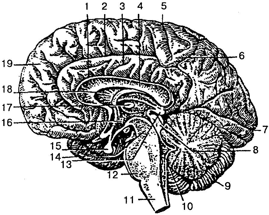
Medial surface of the cerebral hemisphere. All of its lobes take part in the formation of the medial surface of the cerebral hemisphere, except for the insular ( rice. 68). Sulcus of the corpus callosum goes around it from above, separating the corpus callosum from cingulate cortex, goes down and forward and continues in hippocampal sulcus. Passes over the cingulate gyrus cingulate groove, which begins anteriorly and inferiorly from the beak of the corpus callosum, rises upward, turns back, running parallel to the groove of the corpus callosum. At the level of its ridge, a marginal part extends upward from the cingulate groove, which limits the pericentral lobule behind, and the precuneus in front, the groove itself continues into the subparietal groove. Inferiorly and posteriorly through the isthmus, the cingulate gyrus passes into parahippocampal gyrus which ends in front crochet and bounded from above hippocampal sulcus. The cingulate parahippocampal gyrus and the isthmus are combined under the name vaulted. Located deep in the hippocampal sulcus dentate gyrus. The medial surface of the occipital lobe is separated parieto-occipital sulcus from the parietal lobe. From the posterior pole of the hemisphere to the isthmus of the vaulted gyrus passes calcarine groove, which limits from above lingual gyrus. Between the parieto-occipital sulcus in front and the calcarine sulcus behind is located wedge, facing an acute angle anteriorly.

Rice. 68. Brain. Medial surface of the hemisphere. 1 - paracentral lobule, 2 - cingulate gyrus, 3 - cingulate sulcus, 4 - septum pellucidum, 5 - superior frontal sulcus, 6 - interthalamic fusion, 7 - anterior commissure, 8 - thalamus, 9 - hypothalamus, 10 - quadrigeminal, 11 - optic chiasm, 12 – mastoid body, 13 – pituitary gland, 14 – IV ventricle, 15 – pons, 16 – reticular formation, 17 – medulla oblongata, 18 – cerebellar vermis, 19 – occipital lobe, 20 – calcarine groove, 21 – cerebral peduncle , 22 – wedge, 23 – midbrain aqueduct, 24 – occipitotemporal sulcus, 25 – choroid plexus, 26 – fornix, 27 – precuneus, 28 – corpus callosum
Inferior surface of the cerebral hemisphere has the most complex terrain ( rice. 69). In front is the lower surface of the frontal lobe, behind it is the temporal pole and the lower surface of the temporal and occipital lobe y, between which there is no clear boundary. On the lower surface of the frontal lobe, the olfactory groove runs parallel to the longitudinal fissure, to which the olfactory bulb and the olfactory tract, which continues into the olfactory triangle, are adjacent below. Between the longitudinal fissure and the olfactory sulcus there is a straight gyrus. Lateral to the olfactory sulcus lie the orbital gyri. The lingual gyrus of the occipital lobe is bounded by the collateral sulcus, which passes to the inferior surface of the temporal lobe, dividing the parahippocampal and medial occipitotemporal gyri. Anterior to the collateral groove is the nasal groove, which borders the anterior end of the parahippocampal gyrus uncus.

Rice. 69. Control of organs by cranial nerves, diagram. I – olfactory nerve; II – optic nerve; III – oculomotor nerve; IV – trochlear nerve; V – trigeminal nerve; VI – abducens nerve; VII – facial nerve; VIII – vestibulocochlear nerve; IX – glossopharyngeal nerve; X – vagus nerve; XI – accessory nerve; XII – hypoglossal nerve
The structure of the cerebral cortex. The cerebral cortex is formed by gray matter, which lies along the periphery (on the surface) of the cerebral hemispheres. The thickness of the cortex of different parts of the hemispheres ranges from 1.3 to 5 mm. For the first time, Kyiv scientist V.A. Betz showed that the structure and relative position of neurons are not the same in different parts of the cortex, which determines the neurocytoarchitecture of the cortex. Cells of more or less the same structure are arranged in the form individual layers(records). In the neocortex, most neurons form six laminae. In different sections, their thickness, the nature of the boundaries, the size of the cells, their number, etc. vary.
Outside is the first molecular plate, in which small multipolar associative neurons and many fibers of the processes of neurons in the underlying layers lie. Second outer granular plate formed by many small multipolar neurons. The third, the widest, pyramidal plate contains pyramidal-shaped neurons, the bodies of which increase in the direction from top to bottom. Fourth internal granular plate formed by small stellate-shaped neurons. In the fifth internal pyramidal plate, which is most well developed in the precentral gyrus, contains very large (up to 125 µm) pyramidal cells, discovered by V.A. Betz in 1874. Neurons are located in the sixth multiform plate various shapes and sizes.
The number of neurons in the cortex reaches 10–14 billion. In addition to nerve cells, each cell plate contains nerve fibers. K. Brodman in 1903–1909 identified 52 cytoarchitectonic fields in the cortex. O. Vogt and C. Vogt(1919–1920), taking into account the fiber structure, described 150 myeloarchitectonic areas in the cerebral cortex.
Localization of functions in the cerebral cortex. In the cerebral cortex, all stimuli that come from the external and internal environment are analyzed.
In the cortex postcentral gyrus and superior parietal lobule lie nuclei of the cortical analyzer of proprioceptive and general sensitivity(temperature, pain, tactile) of the opposite half of the body. In this case, the cortical ends of the sensitivity analyzer of the lower extremities and lower parts of the body are located closer to the longitudinal fissure of the brain, and the receptor fields of the upper parts of the body and head are projected lowest at the lateral sulcus ( rice. 70A). Motor analyzer core is located mainly in precentral gyrus And paracentral lobule on the medial surface of the hemisphere (“motor cortex”). In the upper parts of the precentral gyrus and paracentral lobule, the motor centers of the muscles of the lower extremities and the lowermost parts of the body are located. In the lower part, near the lateral groove, there are centers that regulate the activity of the muscles of the face and head ( rice. 70B). The motor areas of each hemisphere are connected to the skeletal muscles of the opposite side of the body. The muscles of the limbs are connected in isolation to one of the hemispheres; the muscles of the trunk, larynx and pharynx are connected to the motor areas of both hemispheres. In both centers described, the size of the projection zones of various organs depends not on their size, but on their functional significance. Thus, the zones of the hand in the cerebral hemisphere cortex are much larger than the zones of the trunk and lower limbs combined.
On the surface of the middle part of the temporal gyrus facing the insula there is core of the auditory analyzer. Conducting pathways from the hearing organ receptors on both the left and right sides approach each hemisphere.
Visual analyzer core located on the medial surface of the occipital lobe of the cerebral hemisphere on both sides (“along the banks”) of the calcarine groove. The nucleus of the visual analyzer of the right hemisphere is connected by pathways with the lateral half of the retina of the right eye and the medial half of the retina of the left eye; left with the lateral half of the retina of the left eye and the medial half of the retina of the right eye.

Rice. 70. Location of cortical centers. A – Cortical center of general sensitivity (sensitive “homunculus”) (from V. Penfield and I. Rasmussen). Images of a cross section of the brain (at the level of the postcentral gyrus) and associated symbols show the spatial representation of the body surface in the cerebral cortex. B – Motor area of the cortex (motor “homunculus”; (from V. Pentfield and I. Rasmussen). The image of the motor “homunculus” reflects the relative sizes of the areas of representation of individual parts of the body in the cortex of the precentral gyrus of the cerebrum
Cortical end of the olfactory analyzer - it is a hook and also old and ancient bark. The old cortex is located in the area of the hippocampus and the dentate gyrus, the ancient cortex is located in the area of the anterior perforated space, the septum pellucidum and the olfactory gyrus. Due to the close location of the nuclei of the olfactory and gustatory analyzers, the senses of smell and taste are closely related. The nuclei of the taste and olfactory analyzers of both hemispheres are connected by pathways with receptors on both the left and right sides.
The described cortical ends of the analyzers carry out the analysis and synthesis of signals coming from the external and internal environment of the body, components first signaling system reality (I.P. Pavlov). Unlike the first one, second signaling system exists only in humans and is closely related to the development of articulate speech.
Speech and thinking in humans are carried out with the participation of the entire cerebral cortex. At the same time, there are zones in the cortex that are centers of a number of special functions related to speech. Motor analyzers of oral and written speech are located in areas of the frontal lobe cortex adjacent to the precentral gyrus near the nucleus of the motor analyzer. Analyzers for visual and auditory speech perception are located near the cores of the visual and hearing analyzers. In this case, speech analyzers in right-handers are localized only in the left hemisphere, and in left-handers only in the right.
Basal (subcortical central) nuclei and white matter telencephalon. In the thickness white matter Each cerebral hemisphere has accumulations of gray matter that form separate nuclei that lie closer to the base of the brain. These kernels are called basal(subcortical central). These include striatum, fence And almond-shaped body. The nuclei of the striatum form the striopallidal system, which, in turn, belongs to the extrapyramidal system involved in the control of movements and the regulation of muscle tone.
To the white matter of the hemisphere include the internal capsule and fibers passing through the cerebral commissures (corpus callosum, anterior commissure, fornix commissure) and heading to the cortex and basal ganglia; the fornix, as well as systems of fibers connecting areas of the cortex and subcortical centers within one half of the brain (hemisphere).
Lateral ventricle. The cavities of the cerebral hemispheres are the lateral ventricles (I and II), located in the thickness of the white matter under the corpus callosum. Each ventricle consists of four parts: the anterior horn lies in the frontal lobe, the central part in the parietal lobe, the posterior horn in the occipital lobe, and the inferior horn in the temporal lobe.
diencephalon, located under the corpus callosum, consists of the thalamus, epithalamus, metathalamus and hypothalamus. Thalamus(visual thalamus) paired, formed mainly by gray matter, is the subcortical center of all types of sensitivity. The medial surface of the right and left thalamus, facing each other, forms the lateral walls of the cavity of the diencephalon of the third ventricle. Epithalamus includes the pineal body (epiphysis), leashes and triangles of leashes. The pineal body, which is an endocrine gland, is suspended, as it were, on two leashes connected to each other soldering and connected to the thalamus through leash triangles. The triangles of the leashes contain nuclei related to the olfactory analyzer. Metathalamus formed by paired medial and lateral geniculate bodies lying behind each thalamus. Medial geniculate body along with the lower colliculi of the plate of the roof of the midbrain (quadrigeminal) - subcortical center of the auditory analyzer. Lateral geniculate body together with the superior colliculi of the plate of the roof of the midbrain is subcortical center of the visual analyzer. The nuclei of the geniculate bodies are connected with the cortical centers of the visual and auditory analyzers.
Hypothalamus located anterior to the cerebral peduncles and includes a number of structures: located anteriorly visual part(optic chiasm, optic tract, gray tubercle, infundibulum, neurohypophysis) and olfactory part(mastoid bodies and the subthalamic region itself, the subthalamus). The functional role of the hypothalamus is very large (see section “Endocrine glands”, p. XX). It contains the centers of the autonomic part of the nervous system. The medial hypothalamus contains neurons that perceive all changes occurring in the blood and cerebrospinal fluid (temperature, composition, hormone content, etc.). The medial hypothalamus is also connected to the lateral hypothalamus. The latter does not have nuclei, but has bilateral connections with the overlying and underlying parts of the brain. The medial hypothalamus is the link between the nervous and endocrine systems. IN last years Enkephalins and endorphins, which have a morphine-like effect, are isolated from the hypothalamus. They are involved in the regulation of behavior and vegetative processes. The hypothalamus regulates all body functions except heart rate, blood pressure and spontaneous respiratory movements, which are regulated by the medulla oblongata.
Mastoid bodies, formed by gray matter covered with a thin layer of white, are the subcortical centers of the olfactory analyzer. Located anterior to the mastoid bodies gray bump, which contains the nuclei of the autonomic nervous system. They also influence a person's emotional reactions. The part of the diencephalon located below the thalamus and separated from it by the hypothalamic groove makes up the hypothalamus itself. The coverings of the cerebral peduncles continue here, the red nuclei and the black substance of the midbrain end here.
Cavity of the diencephalon - III ventricle- is a narrow, slit-like space located in the sagittal plane, limited laterally by the medial surfaces of the thalamus, below by the hypothalamus, above by the fornix, above which the corpus callosum is located. The cavity of the third ventricle passes posteriorly into the midbrain aqueduct, and in front on the sides through the interventricular foramina communicates with the lateral ventricles.
TO midbrain include the cerebral peduncles and the roof of the midbrain. Legs brain - these are white round (rather thick) cords emerging from the pons and heading forward to the cerebral hemispheres. Each leg consists of a tire and a base, the boundary between them is black matter(color depends on the abundance of melanin in its nerve cells), relating to the extrapyramidal system, which is involved in maintaining muscle tone and automatically regulates muscle function. Base of the leg formed by nerve fibers running from the cerebral cortex to the spinal and medulla oblongata and the pons. Tegmentum of the cerebral peduncles contains mainly ascending fibers heading to the thalamus, among which the nuclei lie. The largest are red kernels, from which the motor red nucleus-spinal tract begins. In addition, the tire contains reticular formation and the nucleus of the dorsal longitudinal fasciculus (intermediate nucleus).
IN roof of the midbrain differentiate roof plate(quadrigeminal), consisting of four whitish hillocks, two upper (subcortical centers of the visual analyzer) and two lower (subcortical centers of the auditory analyzer). The pineal body lies in the depression between the superior colliculi. The quadrigeminal region is a reflex center for various types of movements that arise mainly under the influence of visual and auditory stimuli. From the nuclei of these hillocks a conducting path originates, ending on the cells of the anterior horns of the spinal cord.
Midbrain plumbing(Aqueduct of Sylvius) is a narrow canal (2 cm long) that connects the III and IV ventricles. Around the water supply is located central gray matter, which contains the reticular formation, nuclei of the III and IV pairs of cranial nerves and other nuclei.
TO hindbrain include the pons, located ventrally, and the cerebellum lying behind the pons. Bridge(Varoliev pons), well developed in humans, looks like a lying transversely thickened ridge, from the lateral side of which extends to the right and left middle cerebellar peduncles. The posterior surface of the pons, covered by the cerebellum, participates in the formation of the rhomboid fossa, the anterior surface (adjacent to the base of the skull) borders the medulla oblongata below and the cerebral peduncles above. The pons consists of many nerve fibers that form pathways and connect the cerebral cortex with the spinal cord and the cerebellar cortex. Between the fibers lie the reticular formation, the nuclei of the V, VI, VII, VIII pairs of cranial nerves.
Cerebellum plays a major role in maintaining body balance and coordination of movements. The cerebellum is well developed in humans due to upright posture and labor activity hands, especially developed cerebellar hemispheres. The cerebellum has two hemispheres and an unpaired median part - worm. The surfaces of the hemispheres and the vermis are separated by transverse parallel grooves, between which there are narrow, long layers of the cerebellum. Due to this, its surface area in an adult is on average 850 cm 2, and its mass is 120–160 g. The cerebellum consists of gray and white matter. The white matter, penetrating between the gray matter, seems to branch, forming white stripes, resembling in the median section the figure of a branching tree - the “tree of life” of the cerebellum ( see fig. 68). The cerebellar cortex consists of gray matter 1–2.5 mm thick. In addition, in the thickness of the white matter there are accumulations of gray, four pairs of nuclei. The nerve fibers connecting the cerebellum with other parts form three pairs cerebellar peduncles: inferior directed to the medulla oblongata, average to the bridge, upper to the quadrigeminal.
The cerebellar cortex has three layers: the outer molecular layer, the middle layer of piriform neurons (ganglionic) and the inner granular layer. The molecular and granular layers contain mainly small neurons. Large piriform neurons (Purkinje cells) measuring up to 40 microns, located in the middle layer in one row, are efferent neurons of the cerebellar cortex. Their axons, extending from the base of the bodies, form the initial link of the efferent pathways. They are directed to the neurons of the cerebellar nuclei, and the dendrites are located in the superficial molecular layer. The remaining neurons of the cerebellar cortex are intercalary (associative), they transmit nerve impulses piriform neurons.
ATTENTION
All nerve impulses entering the cerebellar cortex reach the piriform neurons.
By the time of birth, the cerebellum is less developed than the telencephalon (especially the hemisphere), but in the first year of life it develops faster than other parts of the brain. A marked enlargement of the cerebellum is observed between the fifth and eleventh months of life, when the child learns to sit and walk.
Medulla is a direct continuation of the spinal cord. Its length is about 25 mm, its shape approaches a truncated cone, with the base facing upward. Front surface divided anterior median fissure, on the sides of which are located pyramids, formed by partially intersecting bundles of nerve fibers of the pyramidal pathways. The posterior surface of the medulla oblongata is divided posterior median sulcus, on the sides of it there are continuations of the posterior cords of the spinal cord, which diverge upward, turning into inferior cerebellar peduncles. The latter limit from below rhomboid fossa. The medulla oblongata is built of white and gray matter, the latter is represented by the nuclei of the IX–XII pairs of cranial nerves, olives, centers of respiration and circulation, and the reticular formation. White matter is formed by long and short fibers that make up the corresponding pathways. Centers of the medulla oblongata - blood pressure heart rate and spontaneous respiratory movements. Fibers of the pyramidal tracts connect the cerebral cortex with the nuclei of the cranial nerves and the anterior horns of the spinal cord.
Reticular formation is a collection of cells, cell clusters and nerve fibers located in the brain stem (medulla oblongata, pons and midbrain) and forming a network. The reticular formation is connected to all sense organs, motor and sensory areas of the cerebral cortex, the thalamus and hypothalamus, and the spinal cord. The reticular form regulates the level of excitability and tone of various parts of the central nervous system, including the cerebral cortex, and is involved in the regulation of consciousness, emotions, sleep and wakefulness, autonomic functions, and purposeful movements.
IV ventricle – this is the cavity of the rhombencephalon, continuing downwards into center channel spinal cord. The bottom of the fourth ventricle due to its shape is called rhomboid fossa. It is formed by the posterior surfaces of the medulla oblongata and the pons, the upper sides of the fossa are the upper, and the lower are the inferior cerebellar peduncles. In the thickness of the rhomboid fossa lie the nuclei of the V, VI, VII, VIII, IX, X, XI and XII pairs of cranial nerves.
From the book Marijuana: Myths and Facts by Lynn Zimmer7. Marijuana and the brain MYTHMarijuana kills brain cells. Long-term use of marijuana causes permanent damage to the structure and function of the brain, leading to memory loss, cognitive impairment, personality disorders and decline.
From the book Nervous Diseases: Lecture Notes author A. A. Drozdov1. The brain and its structure The brain consists of two hemispheres, which are separated from each other by a deep groove reaching the corpus callosum. The corpus callosum is a massive layer of nerve fibers that connects both hemispheres of the brain.
From book Newest victories medicine by Hugo GlaserChapter VI Brain and nerves Advances in brain surgery Many thousands of years ago, mankind knew about the operation of craniotomy. During excavations of ancient graves and burials in deep layers of the earth, skulls with well-healed
From the book Histology author V. Yu. Barsukov23. Nervous system. Brain The brain also contains gray and white matter, but the distribution of these two components is more complex here than in the spinal cord. Brain stem. All nuclei of the gray matter of the brainstem consist of multipolar nerve cells. On
From the book Neurology and Neurosurgery author Evgeniy Ivanovich Gusev1.4. Brain 1.4.1. Medulla oblongata The medulla oblongata is a continuation of the spinal cord. The spinal cord passes into the medulla oblongata gradually, without a sharp boundary. The conventional boundary of the transition of the spinal cord into the medulla oblongata is the decussation
From the book Kinesitherapy of Joints and Spine author Leonid Vitalievich RudnitskyBRAIN The brain is divided into gray matter and white matter. Gray matter is a collection of nerve cells that is located in the cerebral cortex. Each area of the cortex is a nerve center that controls a particular function
From the book Homeopathic treatment of cats and dogs by Don Hamilton From the book Spinal Hernia. Non-surgical treatment and prevention author Alexey Viktorovich SadovBrain The brain is divided into gray and white matter. Gray matter is a collection of nerve cells that is located in the cerebral cortex. Each section of the cortex is a nerve center that controls one or another function of the body. From nerve
From the book Alcoholism author Alexander Vitalievich MelnikovBrain Damage to the brain drinking people is determined by two factors: 1) alcohol has a neurotoxic effect, that is, it directly causes the death of cells in the cerebral cortex; 2) disruption of brain functions is caused by a lack of
From the book Healthy to Death. The result of the study of the main ideas about healthy way life author AJ JacobsChapter 11 Brain Goal: become smarter There has never been a better time for fools in history. Never before have so many people believed that with hard work and the right techniques, you can improve your brain and become smarter. For decades, it was believed that intelligence was given by nature,
From the book Five Steps to Immortality author Boris Vasilievich BolotovBrain Double vision, speech retardation, impaired motor coordination, epilepsy, parkinsonism, multiple sclerosis, schizophrenia, mottled skin coloration. Source plant material: peony, cocklebur (non-rebe), mandrake, poppy, hemp, tobacco,
From the book A Healthy Man in Your Home author Elena Yurievna ZigalovaBrain The brain is located in the cranial cavity. The brain mass does not exceed 2% of the total body mass. On average, the brain of an adult male weighs 1375–1400 g. Moreover, the relative weight of the brain of men is less than that of women. So, in men, per 1 kg of body weight
Second higher education"Psychology" in MBA format
item:Anatomy and evolution of the human nervous system.
Manual "Anatomy of the central nervous system"
2.1. General diagram of the structure of the central nervous system
2.2. Brain cavities and cerebrospinal fluid
2.3. Meninges
2.1. General diagram of the structure of the central nervous system
In the nervous system there are central and peripheral nervous system.
The peripheral nervous system is represented by:
roots of the spinal cord,
nerve plexuses,
nerve ganglia (ganglia),
nerves
peripheral nerve endings (Fig. 2.1).
Rice. 2.1. Components of the peripheral nervous system :
In its turn, nerve endings can be:
A) efferent
(motor), which transmit excitation from nerves to muscles and glands;
b) afferent
(sensitive), transmitting information from receptors to the central nervous system.
The human central nervous system consists of the brain and spinal cord.
Spinal cord It is a tube with a small channel in the middle, surrounded by neurons and their processes.
Brain is an extension of the spinal cord.
In the distant ancestors of chordates (for example, the lancelet), the neural tube has the same diameter throughout, and the brain is practically absent. In fish, the brain is already well developed, and with each stage of evolution it increases. The brain reaches its highest development in a person who has the most big indicator cephalization (the ratio of brain mass to body mass) among all other living beings.
Macroscopically (with the naked eye) on a section of the brain one can distinguish white and gray matter.
White matter
represents bundles of nerve fibers and forms pathways. Since most of the long nerve processes are covered with a layer of white fat-like substance (myelin), their clusters have White color.
Gray matter
- these are the bodies of neurons that form nerve centers. Gray matter in the central nervous system forms two types of clusters (structures): nuclear structures
(nuclei of the spinal cord, brainstem and cerebral hemispheres), in which cells lie in close groups, and screen structures
(cerebral cortex and cerebellum), in which cells lie in layers.
Brain
lies in the cranial cavity. The topographic boundary with the spinal cord is a plane passing through the lower edge of the foramen magnum. The average brain mass is 1400 g with individual variations from 1100 to 2000. There is no clear connection between brain mass and a person’s intellectual abilities. Thus, the brain of I. S. Turgenev reached a mass of almost 2 kg, and French writer Anatole France weighed a little more than one kilogram. However, their contribution to world literature equal size
Anatomically in the brain one can distinguish the hemispheres, the brainstem and the cerebellum (small brain).
Trunk includes medulla oblongata, pons, midbrain and diencephalon (Fig. 2.2).
Rice. 2.2. Anatomical parts of the brain
There is another classification of brain regions, which focuses on the developmental features of a particular region (during the process of ontogenesis). If the parts of the brain are distinguished based on the processes of embryonic development (in accordance with the stage of the three brain vesicles), then the brain can be divided into forebrain, midbrain and hindbrain (diamond-shaped). In accordance with this approach, the forebrain includes the cerebral hemispheres and diencephalon, the midbrain includes the midbrain, and the rhomboid (developing from the hindbrain vesicle) includes the medulla oblongata, hindbrain and isthmus of the rhombencephalon (Fig. 2.3).
Rice. 2.3. Ontogenetic classification of brain regions
Left and right hemisphere The telencephalon is separated by a longitudinal fissure, the bottom of which is the corpus callosum. They are separated from the cerebellum by a transverse fissure. The entire surface of the hemispheres is covered with grooves and convolutions, the largest of which is the lateral, or Sylvian, which separates the frontal lobe of the hemispheres from the temporal.
A sagittal section of the brain shows the medial surface of the cerebral hemispheres, the structures of the brain stem and cerebellum (Fig. 2.4).
Rice. 2.4. Sagittal section of the human brain:
1 - forebrain hemisphere;
2 - cerebellum;
3 - medulla oblongata;
4 - bridge;
5 - midbrain;
6 - diencephalon;
7 - corpus callosum
12 pairs of cranial nerves depart from the brain, innervating mainly the head, a number of muscles of the neck and back of the head, and also providing parasympathetic innervation internal organs. 31 pairs of spinal nerves depart from the spinal cord, innervating the torso and internal organs.
The cerebral cortex is separated by a groove from the corpus callosum. The corpus callosum is a large commissure of the brain and has a fibrous structure. Under the corpus callosum there is a thin white stripe - the fornix.
2.2. Brain cavities and cerebrospinal fluid
During embryonic development, the cavities of the brain vesicles are transformed into the ventricles of the brain. The first and second ventricles are located in the left and right hemispheres, respectively, the third ventricle is located in the diencephalon, and the fourth ventricle is located in the rhombencephalon. The third and fourth ventricles are connected by the aqueduct of Sylvius, which runs in the midbrain. The cavities of the brain are filled with cerebrospinal fluid (CSF). They communicate with each other, as well as with the spinal canal and the nodiautinal space (the space under one of the membranes of the brain) (Fig. 2.5).
Rice. 2.5. Diagram of brain cavities
Cerebrospinal fluid is produced by the choroid plexuses of the ventricles of the brain, which have a glandular structure, and is absorbed by the veins of the pia mater of the brain. The processes of formation and absorption of cerebrospinal fluid occur continuously, providing a 4-5-fold exchange of cerebrospinal fluid within one day. In the cranial cavity there is a relative insufficiency of cerebrospinal fluid absorption (i.e., less cerebrospinal fluid is absorbed than is produced), and in the intravertebral canal a relative insufficiency of cerebrospinal fluid production predominates (less cerebrospinal fluid is produced than is absorbed). When the cerebrospinal fluid dynamics between the brain and spinal cord is disrupted, excessive accumulation of cerebrospinal fluid develops in the cranial cavity, and in the subarachnoid space of the spinal cord the fluid is quickly absorbed and concentrated.
The circulation of cerebrospinal fluid depends on the pulsation of the blood vessels of the brain, breathing, head movements, the intensity of the formation and absorption of the cerebrospinal fluid itself.
From the lateral ventricles of the brain, where, we repeat, the formation of cerebrospinal fluid dominates over its absorption, cerebrospinal fluid enters the third ventricle of the brain and further, along the brain aqueduct, into the fourth ventricle, from where, through the foramina of Luschka, the cerebrospinal fluid enters the cistern magna and the external subarachnoid space of the brain , central canal and subarachnoid space of the spinal cord and into the spinal cord cistern terminale.
Functions of cerebrospinal fluid
. Mechanical protection of the brain.
. Damping changes in osmotic pressure.
. Maintaining trophic and metabolic processes between blood and brain.
2.3. Meninges
The brain and spinal cord are surrounded by membranes that perform protective functions.
There are dura mater, arachnoid mater and pia mater.
Solid
The meninges are located most superficially.
Arachnoid (arachnoid)
the shell occupies a middle position.
Soft
the membrane is directly adjacent to the surface of the brain. It seems to “envelop the brain”, entering all the grooves, and is separated from the arachnoid membrane by the subarachnoid space filled with cerebrospinal fluid. Strands and plates are stretched between the soft and arachnoid membranes, thus the vessels passing through them are “suspended”. The subarochnoid space forms expansions, or cisterns, filled with cerebrospinal fluid. There are the cerebellopontine (larger) cistern, interpeduncular cistern, chiasmal cistern, and terminal cistern (spinal cord).
The arachnoid is separated from the nerdon of the meninges by the capillary subral space. It consists of two leaves. The outer leaf is attached to the skull from the inside and sends out the internal canal of the spine, making up their periosteum. The inner leaf is fused with the outer one (forming at the fusion sites the so-called cerebral sinuses of the bed for the outflow of venous blood from the brain and head). Between the outer layer of the skull bones and the vertebrae is the epidural space.
The brain is part of the central nervous system that is located inside the skull. The brain controls all functions of the body, including the rhythm of heart contractions, the ability to walk and run, and the occurrence of our thoughts and emotions.
The brain consists of three main sections - the hindbrain, midbrain and forebrain. The forebrain is divided into two halves - the left and right hemispheres of the brain.
Cerebral hemispheres
The cerebral hemispheres make up the largest part of the forebrain. Their outer surface forms a folded system of convolutions and grooves, which significantly increases the surface area. Most of the surface of the brain is hidden deep in the sulci. Each hemisphere is divided into the frontal, parietal, occipital and temporal lobes, named after the bones of the skull closest to them. The corpus callosum connects both hemispheres - a large bundle of fibers deep in the longitudinal fissure of the brain.Gray and white matter of the brain
The hemispheres consist of an outer cortex of gray matter and an inner mass of white matter.The gray matter of the brain contains the bodies of nerve cells and forms the cerebral cortex, the cerebellar hemispheres and a group of subcortical nuclei.
The white matter consists of nerve fibers and is located under the cortex. Nerve fibers provide communication between the halves of the brain, as well as with the spinal cord and the entire body.
Furrows and convolutions
The central sulcus is located between the longitudinal and lateral sulci and forms the boundary between the frontal and parietal lobes. The precentral gyrus runs parallel and anterior to the central sulcus and contains the primary motor cortex, which is responsible for voluntary movements. The postcentral gyrus contains the primary somatosensory cortex, which perceives sensory sensations. The parieto-occipital sulcus (on the inner surface of both hemispheres) separates the parietal and occipital lobes.The calcarine sulcus marks the location of the primary visual cortex, where visual information is perceived. The primary auditory cortex is located posterior to the lateral sulcus.
On the inner surface of the temporal lobe is the primary olfactory cortex, where smell analysis occurs. Inward to the parahippocampal gyrus lies the hippocampus, which is part of the limbic system and is involved in memory formation. The areas responsible for speech are located in the dominant hemisphere (usually the left) of each individual. The motor speech center (Broca's area) is located in the posterior parts of the inferior frontal gyrus; it is necessary in the process of speech formation.
Inside the brain
A section of the brain along the midline between the two cerebral hemispheres shows the major structures that control numerous body functions. While some areas of the brain process sensory and motor information, others control speech and sleep.Speech, thinking and motor activity
The sensory speech center (Wernicke's area) lies behind the primary auditory cortex and is necessary for understanding speech. The prefrontal cortex is responsible for cognitive functions higher order, including abstract thinking, social behavior and decision making. Within the white matter of the cerebral hemispheres are areas of gray matter known as the basal ganglia. This group of structures regulates different kinds motor activity.Diencephalon
The diencephalon is the middle part of the forebrain and includes structures bordering the third ventricle.These include: the thalamus, hypothalamus, as well as the epithalamus and subthalamus. The thalamus is the last way station for information from the brainstem and spinal cord before it reaches the cortex. The hypothalamus lies beneath the thalamus in the lower part of the diencephalon. It is responsible for various mechanisms of homeostasis (maintenance of life), and also controls the pituitary gland, which descends from the base of the hypothalamus. The anterior lobe of the pituitary gland secretes substances that regulate the activity of the thyroid gland, adrenal glands and ovaries and produces growth factors. The posterior lobe secretes hormones that increase blood pressure, decrease urine production, and cause uterine contractions.
The hypothalamus also influences the sympathetic and parasympathetic nervous systems and regulates body temperature, appetite, and sleep-wake patterns. The epithalamus is a relatively small part of the posterior part of the diencephalon, which includes the pineal gland (epiphysis), which synthesizes melatonin.
The subthalamus is located inferior to the thalamus next to the hypothalamus. Contains the subthalamic nucleus, which is involved in the regulation of movements.
Brain stem and cerebellum
The posterior part of the diencephalon is related to the midbrain, followed by the pons and medulla oblongata, which are related to the hindbrain. The midbrain and hindbrain contain nerve fibers that connect the cerebral hemispheres with the nuclei of the cranial nerves, with underlying centers in the brain stem and with the spinal cord. The midbrain and hindbrain also contain cranial nerve nuclei.Most of the reticular formation - the system of nerve pathways - lies in the midbrain and hindbrain. This system contains vital centers: respiratory, cardiac and vasomotor (vasomotor).
The cerebellum lies behind the hindbrain and is connected to it through three pairs of cerebral peduncles. Connections to the rest of the brain and spinal cord are made through these peduncles. The cerebellum functions at an unconscious level, coordinating movements initiated in other areas of the brain, and also provides balance, maintaining posture and muscle tone.
The human body. Outside and inside. №14 2008
Brain(encephalon) with its surrounding membranes is located in the cavity of the cerebral part of the skull.
The upper convex surface of the brain corresponds in its shape to the inner surface of the cranial vault, and the lower, flatter, with complex relief, corresponds to the inner base of the skull.
The weight of the adult human brain ranges from 1100 to 2000 g; in men, on average, it is about 1394 g, and in women, 1245 g. After 60 years, the mass and volume of the brain decrease slightly.
The largest parts of the brain are the cerebral hemispheres, the cerebellum and the brain stem.
In an adult, the cerebral hemispheres are the largest and functionally important part of the central nervous system; they cover other brain structures. Right and left hemisphere separated from one another by a deep longitudinal fissure reaching the corpus callosum, or large commissure of the brain. The longitudinal fissure flows posteriorly into the transverse fissure of the cerebrum, which separates the hemispheres from the cerebellum.
On the surface of the cerebral hemispheres there are deep and shallow grooves. Deep grooves divide each hemisphere into lobes, and shallow grooves are separated from one another by the convolutions of the cerebrum. The base of the brain is formed by the ventral surfaces of the cerebral hemispheres, the cerebellum, and the ventral sections of the brain stem.
At the base of the brain (Fig. 106), in its anterior part, there are olfactory bulbs, from which extends a large nerve cord - the olfactory tract, which passes into the olfactory triangle. Behind it is a perforated substance formed by arteries penetrating deep into the brain. Inside the anterior perforated substance is the optic chiasm, formed by fibers of the optic nerve, which, partially crossing, emerge from the chiasm in the form of optic tracts. Adjacent to the posterior surface of the optic chiasm is a gray tubercle, the lower sections of which, tapering, form a funnel in which the pituitary gland, an endocrine gland, is located. The gray tubercle is joined by two white spherical elevations - the mastoid bodies. Behind the visual pathways two longitudinal white ridges are visible - peduncles of the brain between them. there are depressions - the interpeduncular fossa, the bottom of which is filled with perforated substance. A little further away there is a wide transverse roller - bridge.
Rice. 106. Base of the brain:
1 - olfactory bulb; 2 - olfactory tract; 3 - anterior perforated substance; 4 - gray tubercle; 5- optic tract; 6- mastoid bodies; 7 - trigeminal node; 8 - posterior perforated substance; 9- bridge; 10- cerebellum; 11- pyramid of the medulla oblongata; 12- olive; 13 - spinal nerves; 14 - hypoglossal nerve; 15 - accessory nerve; 16- nervus vagus; 17- glossopharyngeal nerve; 18- vestibulocochlear nerve; 19 - facial nerve; 20 - abducens nerve; 21 - trigeminal nerve; 22 - trochlear nerve; 23 - oculomotor nerve; 24 - optic nerve; 25 - olfactory nerves
The lateral sections of the pons continue into the cerebellum and form its middle cerebellar peduncles. Below the bridge are the anterior sections of the medulla oblongata, which are represented by medially located pyramids, separated from each other by the anterior median fissure; olives are identified laterally.
A sagittal section of the brain (Fig. 107) shows its various structures - areas of the frontal, parietal and occipital lobes, the corpus callosum, which is separated from them by a corresponding groove. The middle part of the corpus callosum is called the trunk, the anterior part is called the knee. Below, the genu of the corpus callosum becomes thinner, passes into the beak of the corpus callosum, and the posterior sections end in the form of a ridge.
Rice. 107. Brain (sagittal section):
1 - groove of the corpus callosum; 2 - cingulate groove; 3 - cingulate gyrus; 4- corpus callosum; 5- central groove; 6- paracentral lobule; 7- calcarine groove; 8- roof plate (quadrigeminal); 9 - cerebellum; 10 - IV ventricle; 11 - medulla oblongata; 12 - bridge; 13 - pineal body (epiphysis); 14- cerebral peduncles; 15- pituitary; 16- III ventricle; 17-interthalamic fusion; 18 - transparent partition; 19 - superior frontal gyrus
Beneath the corpus callosum is a thin white plate called the body of the fornix, which continues into the column of the fornix. The latter ends with the mastoid body, and behind it passes into the crura of the fornix. A bundle of nerve fibers runs transversely between the columns of the fornix - the anterior commissure of the brain, connecting the cerebral hemispheres. The columns of the fornix surround a thin plate of medulla - the transparent septum. All of the above formations are part of the telencephalon, and the structures located below belong to the brain stem (intermediate, middle, posterior parts of the brain and medulla oblongata). The anterior parts of the brain stem are formed by the visual tuberosities, which are located downward from the body of the fornix and the corpus callosum and behind the columns of the fornix. On the median section, only the medial surface of the posterior thalamus (optic thalamus) is visible, which limits the slit-like, vertically located cavity of the third ventricle. Between the anterior end of the thalamus and the column of the fornix there is an interventricular foramen, through which the lateral ventricle of the hemisphere is connected to the cavity of the third ventricle, in the formation of the bottom of which the optic chiasm, gray tubercle, infundibulum, pituitary gland, and mastoid bodies participate. Above and below the optic thalamus, under the splenium of the corpus callosum, there is the pineal body, the anterior-inferior parts of which are fused by a thin cord running transversely - epithalamic (posterior) commissure. The midbrain aqueduct originates from it below. The optic thalamus, hypothalamus, third ventricle, and pineal body belong to the diencephalon. Below the pineal gland is the roof of the midbrain (plate quadrigeminal), consisting of the superior and inferior colliculi. The ventral plate of the midbrain roof is the cerebral peduncle, separated from the plate by the midbrain aqueduct. The cavities of the third and third ventricles are connected through the midbrain aqueduct. Even more posterior are the pons and cerebellum, which belong to the hindbrain and medulla oblongata. The cavity of these brain organs is the IV ventricle, the bottom of which is formed by the dorsal surfaces of the pons and medulla oblongata, which make up the rhomboid fossa.
The brain is divided into five sections: the medulla oblongata, hindbrain, midbrain, diencephalon and telencephalon.
Medulla(Fig. 108).
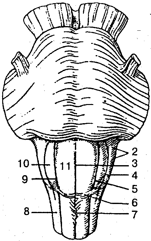
Rice. 108. Medulla oblongata (ventral view):
1 - medulla oblongata; 2 - behind-live field; 3- anterior median fissure; 4 - retroolive groove; 5,9- anterolateral groove; 6- anterior outer arcuate fibers; 7- intersection of pyramids; 8- lateral cord; 10- olive; 11 - pyramid of the medulla oblongata
Located between the hind cord and spinal cord. The lower border of the medulla oblongata corresponds to the level of the foramen magnum, or the exit site of the roots of the first pair of spinal nerves, the upper border runs along the posterior edge of the pons. The length of the medulla oblongata of an adult is on average 25 mm. The upper part of the medulla oblongata, unlike the lower part, has some thickening, which resembles the shape of a cone. The sulci of the brain are a continuation of the sulci of the spinal cord. On the sides of the anterior median fissure on the ventral surface of the medulla oblongata there are convex pyramids, gradually tapering at the bottom, formed by pyramidal tracts, some of the fibers of which form the decussation of the pyramids. Lateral to the pyramid on both sides there are elevations - olives, separated from the pyramid by the anterior lateral groove, from which the roots of the hypoglossal nerve emerge (XII pair of cranial nerves). In the lower part of the dorsal surface of the medulla oblongata there is a dorsal median sulcus, on the sides of which thin and wedge-shaped bundles of the posterior funiculi of the spinal cord end with thickenings, separated from each other by the posterior intermediate sulcus. In the thickenings of the bundles there are corresponding nuclei, from which fibers arise that form the medial loop. The latter forms a decussation at the level of the medulla oblongata. The bundles of this decussation are located in the interolive layer, dorsal to the pyramids. Thin roots of the glossopharyngeal (IX pair), vagus (X pair) and accessory (XI pair) cranial nerves emerge from the posterolateral groove of the medulla oblongata, the nuclei of which lie in the dorsolateral parts of the medulla oblongata. On the dorsal surface, parts of the lateral funiculus expand and, together with fibers from the sphenoid and inferior nuclei, form the inferior cerebellar peduncles, limiting the rhomboid fossa below; the upper part of the dorsal surface participates in the formation of the bottom of the fourth ventricle. The gray matter of the medulla oblongata is represented by clusters of neurons that form the inferior olivary nuclei. Dorsal to the pyramids is the reticular formation, consisting of intertwining fibers - nerve cells.
The medulla oblongata performs reflex and conduction functions. Through the sensitive fibers of the roots of the cranial nerves, it receives information (impulses) from the skin, mucous membranes and organs of the head, as well as from receptors of the larynx, trachea, and internal organs chest(lungs, heart), digestive system. Many simple and complex reflexes are carried out through the medulla oblongata. For example: 1) protective - coughing, sneezing, vomiting, lacrimation, blinking; 2) food - sucking, swallowing, separation of digestive juice; 3) cardiovascular, regulating the activity of the heart and blood vessels; 4) an automatically regulated respiratory center that provides ventilation of the lungs; 5) vestibular nuclei, involved in the implementation of postural adjustment reflexes, in the redistribution of muscle tone.
In addition, paths pass through the medulla oblongata that connect the cerebral cortex, diencephalon and midbrain, cerebellum and spinal cord with a two-way connection.
hindbrain(Fig. 109). Includes the pons and cerebellum.
![]()
Rice. 109. Hindbrain:
1 - cerebellum; 2 - cerebellopontine triangle; 3 - bulbar-pontine sulcus; 4 – basilar groove ; 5 - middle cerebellar peduncle; 6 - bridge
Bridge (pons varoliev) from below it borders with the medulla oblongata, from above it passes into the cerebral peduncles, its lateral sections form the middle cerebellar peduncles (Fig. 110).
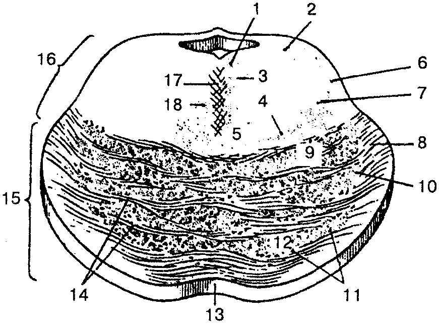
Rice. 110. Cross section of the bridge (diagram):
1 - posterior longitudinal beam; 2 - midbrain tract of the trigeminal nerve; 3- medial longitudinal fasciculus; 4 - medial loop; 5- reticular formation; 6- trigeminal lemniscus (trigeminothalamic tract); 7- spinal loop; 8- pontocerebellar fibers; 9- corticonuclear fibers; 10- corticopontine fibers; 11 - bridge cores; /2-corticospinal fibers; 13 - basilar groove; 14 - cross fibers of the bridge; 15 - anterior (basilar) part of the bridge; 16 - rear part of the axle (axle cover); / 7-suture bridge; 18- tegnospinal tract
From the deep horizontal groove separating the pons from the pyramids of the medulla oblongata, the roots of the abducens nerves (VI pair) emerge, the nuclei of which lie in the dorsal part of the pons. In the lateral part of this groove the roots of the facial (VII pair) and vestibulocochlear (VIII pair) nerves are visible. The continuation of the bridge in the lateral direction forms the middle leg of the bridge. On the ventral surface of the bridge there is a wide but shallow basilar groove; two longitudinal fibers extend from its sides, within which fibers of the pyramidal tracts pass. The dorsal surface of the pons is covered by the cerebellum and is not visible from the outside. It is separated from the dorsal surface of the medulla oblongata by the medullary stripes and together with it participates in the formation of the rhomboid fossa, or the bottom of the fourth ventricle. From the anterolateral sections of the pons, bundles of the trigeminal nerve emerge, the nuclei of which lie in the dorsal part of the pons and medulla oblongata (V pair).
A cross section of the bridge shows a thick bundle of transverse fibers, which belong to the conductive path of the auditory analyzer and form a trapezoidal body. The latter divides the bridge into posterior (tegmental) and anterior (basilar) parts. Between the fibers of this body is the superior olive nucleus. Directly above the trapezoid body lie the fibers of the medial lemniscus, coming from the medulla oblongata; above it is the reticular formation of the bridge. The fibers of the lateral (auditory) lemniscus pass along the side and above the medial lemniscus, and the posterior longitudinal fasciculus lies above the reticular formation.
Cerebellum located posterior to the pons and from the upper part of the medulla oblongata, filling most of the posterior cranial fossa. In the cerebellum, there are superior and inferior surfaces; the boundary between them is the posterior edge of the cerebellum, where a deep horizontal fissure runs. The cerebellum lies on the dorsal surface of the brain stem, covers it from the sides and is connected to its parts with the help of peduncles: the superior cerebellar peduncles connect the cerebellum with the midbrain, the middle peduncles with the pons; lower ones - with the medulla oblongata. The cerebellum is divided into two hemispheres and an unpaired middle part - the cerebellar vermis. On the upper and lower surfaces of the hemispheres and the vermis there are many parallel cerebellar fissures, between which there are long and narrow gyri of the cerebellum.
Groups of gyri, separated by deeper grooves, form the cerebellar lobules. The hemispheres and the cerebellar vermis consist of white matter located medially and a thin layer of gray matter of the cerebellar cortex, which covers the white matter at the periphery. The cerebellar cortex is composed of three layers of nerve cells. When cut through, the white matter of the cerebellum resembles a branched tree, hence its name “tree of life.” In the thickness of the white matter there are separate paired clusters of nerve cells that form the dentate, cork-shaped, spherical nuclei and the tent nucleus.
The hindbrain is a vital part of the nervous system, where the arcs of a number of somatic and autonomic reflexes are closed. With the participation of the hindbrain nuclei, chain reflexes associated with chewing and swallowing are carried out. Many autonomic reflexes of the hindbrain are associated with the function of the digestive tract. These include reflex regulation of the secretion of the salivary glands.
The cerebellum, as a suprasegmental organ, is part of the movement regulation system and performs the following important functions: 1) regulation of posture and muscle tone; 2) sensorimotor coordination of posture and goal-directed movements; 3) coordination of fast, purposeful movements carried out on command from the cerebral cortex.
The main functions of the cerebellum also determine the nature of pathological symptoms when its activity is disrupted. It is known that with partial general damage to the cerebellum, three main symptoms are observed: atony, asthenia and astasia. Atony characterized by weakening muscle tone. In animals after removal of the cerebellum, an initial increase in the tone of the extensor muscles is observed. Their movements are poorly coordinated, sweeping, abrupt, and they are unable to maintain an appropriate posture. Asthenia characterized by weakness and rapid muscle fatigue. Movement tires the animal very much; after walking a few steps, it lies down to rest. The third symptom is astasia - manifests itself in the ability of muscles to perform oscillatory and tremulous movements. Muscle tremors are especially pronounced at the beginning and end of movements, which significantly interferes with purposeful movement.
When the cerebellum is damaged, there is also a symptom ataxia. Patients with this symptom walk with their legs spread wide apart, make unnecessary movements, and sway from side to side. The coordination of voluntary movements in a sitting or lying position changes little.
Midbrain(Fig. 111).

Rice. 111. Midbrain and rhomboid fossa:
1 - roof plate (quadrigeminal); 2 - superior cerebellar peduncle; 3 - loop triangle; 4 - lower colliculus; 5- superior colliculus; 6- handle of the inferior colliculus; 7- handle of the superior colliculus
It contains the roof and legs of the brain. The cavity of the midbrain is the cerebral aqueduct. The lower border of the midbrain on its ventral surface is the anterior edge of the pons, the upper border is the optic tract and the level of the mammillary bodies.
Roof of the midbrain It is a quadrigeminal plate and is located above the cerebral aqueduct. It consists of four elevations ~ mounds, which look like hemispheres, separated from one another by perpendicular grooves. The pineal body is located in the longitudinal groove. A transverse groove separates the pair of superior colliculi from the lower ones. The thickness of the mounds is gray matter.
From each hillock, a thickening extends in the lateral direction - the handle of the hillock, which ends in the geniculate bodies of the diencephalon. In humans, the superior colliculus of the roof of the midbrain (tetracollis) and the lateral geniculate body perform the function of subcortical visual centers. The inferior colliculus and medial geniculate body are subcortical auditory centers.
At the base of the brain, two thick white diverging bundles are clearly visible, extending into the tissue of the cerebral hemispheres. This peduncles of the brain. The depression between them is called the interpeduncular fossa. The roots of the oculomotor nerves (III pair) emerge from it. On a cross section of the midbrain, the black substance is clearly distinguished by its dark color (due to the pigment in the cells - melanin). It extends in the cerebral peduncle from the pons to the diencephalon. The substantia nigra divides the cerebral peduncle into two sections: the posterior - the tegmentum of the brain and the anterior - the base of the cerebral peduncle. In the tegmentum of the midbrain there are ascending pathways and the nuclei of the midbrain lie. The largest tegmental nucleus in a section of the midbrain is red core. It is located slightly above the substantia nigra, has an oblong shape and extends from the level of the inferior colliculus to the hypothalamus.
Red core - one of the central coordination formations of the extrapyramidal system.
Aqueduct of the midbrain (Sylvian aqueduct) - a narrow channel about 1.5 cm long; connects the cavity of the third ventricle with the fourth and contains cerebrospinal fluid. Around the midbrain aqueduct is the central gray matter, in which the nuclei of the III and IV pairs of cranial nerves are located.
The functional significance of the midbrain is that the subcortical centers of hearing and vision are located here; nuclei of the cranial nerves, providing innervation to the striated and smooth muscles of the eyeball; nuclei belonging to the extrapyramidal system (substantia nigra, red nucleus), which provide contraction of the body muscles during automatic movements. In addition, descending (motor) and ascending (sensitive) pathways pass through the midbrain. The midbrain area is also the location of the autonomic centers and the reticular formation.
Damage to the midbrain in animals causes impaired muscle tone. This phenomenon is called decerebrate rigidity. This condition is characterized by a sharp increase in the tone of the extensor muscles of the limbs, back and tail. The animal, placed on its paws, maintains a standing position, since flexion in the joints does not occur. Decerebrate rigidity is a reflex state that is maintained by sensory signals from muscle proprioceptors. This condition occurs because, as a result of transection of the brain stem, the red nuclei and reticular formation are separated from the medulla oblongata and spinal cord.
Diencephalon. Located under the corpus callosum and fornix, fused on the sides with the cerebral hemispheres. It is represented by the following sections: 1) the area of the visual buffalo (thalamic area); 2) hypothalamus (subthalamic region); 3) III ventricle.
The thalamic region includes the thalamus (optic thalamus), metathalamus (medial and lateral geniculate bodies) and epithalamus (pineal body, leashes, leash commissures and epithalamic commissure).
Thalamus - paired ovoid-shaped formations located on the sides of the third ventricle. It consists of gray matter, in which separate clusters of nerve cells are distinguished - the nuclei of the thalamus, separated by thin layers of white matter. Currently, there are up to 120 cores that perform various functions. Due to the fact that most of the sensory pathways are switched here, the thalamus is actually a subcortical sensory center, and its pad is a subcortical visual center.
Metathalamus represented by the lateral and medial geniculate bodies - paired formations that connect to the hillocks of the roof of the midbrain with the help of the handles of the superior and inferior colliculi. The lateral geniculate body, together with the superior colliculi of the midbrain, is the subcortical center of vision. The medial geniculate body and the inferior colliculi of the midbrain form the subcortical hearing centers.
Epithalamus unites the pineal body (epiphysis), leashes and triangles of leashes. The anterior sections of the leashes before the entrance to the epiphysis form a commissure of the leashes. In front and below the pineal body there is a bundle of transversely running fibers - the epithalamic commissure. Between the commissure of the leashes and the epithalamic commissure at the base of the pineal body, a shallow depression is formed - the pineal recess.
Hypothalamus forms the lower parts of the diencephalon, participates in the formation of the bottom of the third ventricle. The hypothalamus includes the optic chiasm, optic tract, mammillary bodies, gray tubercle with infundibulum and pituitary gland.
The optic chiasm consists of fibers of the optic nerves (II pair of cranial nerves), partially passing to the opposite side, and resembles a roller, which then continues into the optic tract. Behind the optic chiasm there is a gray tubercle, below which turns into a funnel, which then connects to the pituitary gland. The mastoid bodies are located between the gray tubercle and the posterior perforated substance and consist of white and gray matter. The columns of the arch of the corpus callosum end in the mastoid bodies. The hypothalamus and pituitary gland form a single functional complex, in which the first plays a regulatory role, and the second an effector.
In the hypothalamus, there are three main hypothalamic regions of the accumulation of nerve cells: anterior, posterior and intermediate. Clusters of nerve cells in these areas form more than 30 nuclei of the hypothalamus. The nerve cells of its nuclei have the ability to produce neurohormones (vasopressin, or antidiuretic hormone, oxytocin), which then enter the posterior lobe of the pituitary gland along the branches of the axons of neurosecretory cells and are carried throughout the body by the blood stream. Some nuclei of the hypothalamus produce so-called releasing factors (liberins) and inhibitory factors (statins) that regulate the activity of the adenohypophysis. The latter transmits information further in the form of tropic hormones to the peripheral endocrine glands. The releasing factor promotes the release of thyroid-, luteo-, corticotropin, prolactin, somato- and melanotropin. Statins inhibit the release of the latter two hormones and prolactin. Peptide-like substances enkephalins and endorphins, which have a morphine-like effect, have also been isolated from the hypothalamus. It is believed that these substances are involved in the regulation of behavior and vegetative processes.
The main functions of the thalamus are the integration (unification) of all types of sensitivity, except for smell; comparison of information received on different communication channels and assessment of its biological significance. According to their function, thalamic nuclei are divided into specific, nonspecific, and associative.
In specific nuclei, sensory information is switched from axons of ascending afferent pathways to terminal neurons, the processes of which go to the sensory areas of the cerebral cortex. Damage to these nuclei leads to irreversible loss of certain types of sensitivity. The nonspecific nuclei of the thalamus are connected with the basal ganglia and various parts of the brain; they maintain a certain level of brain excitability necessary for the perception of irritation from environment. Associative nuclei are involved in high integration processes.
In humans, the thalamus plays a significant role in emotional behavior, which is characterized by peculiar facial expressions, gestures, and shifts in the functions of internal organs. At emotional reactions rises arterial pressure, heart rate and breathing speed up, pupils dilate. Damage to the thalamus in humans is accompanied by severe headaches, disturbances in sleep and sensitivity, movement coordination, accuracy, etc.
The hypothalamus is the main subcortical center of the autonomic nervous system, plays big role in maintaining the constancy of the internal environment of the body, ensures the integration of the functions of the autonomic, endocrine and somatic systems. In addition, the hypothalamus is involved in the formation of versatile behavioral reactions, plays a significant role in thermoregulation, determines the correct periodicity of functions associated with reproduction. As a regulatory organ, the hypothalamus is involved in the alternation of sleep and wakefulness, as well as in the regulation of the activity of the pituitary gland, and has a connection with the limbic system.
Finite brain. It consists of two hemispheres of the cerebrum, separated by a longitudinal fissure and connected in it by the corpus callosum, anterior and posterior commissures, and the commissure of the fornix. The telencephalon cavity forms the right and left lateral ventricles, each of them located in its own hemisphere. The cerebral hemisphere consists of the cerebral cortex (cloak) and the underlying white matter and gray matter located in it - the basal ganglia. The border between the telencephalon and the diencephalon is located at the place where the internal capsule is adjacent to the lateral side of the thalamus.
Cerebral hemispheres covered on the outside with a thin plate of gray matter - the cerebral cortex.
The surface area of the cerebral cortex in an adult is on average 220 thousand mm, with the convex parts of the gyri accounting for 1/3, and the lateral and lower walls of the sulci accounting for 2/3 of the total area of the cortex. The cortex contains about 14 billion neurons. There are six layers of nerve cells in the cortex: 1) molecular plate; 2) external granular plate; 3) external pyramidal plate; 4) internal granular plate; 5) internal pyramidal plate; 6) multiform plate. In each layer, in addition to the cells, there are their processes - fibers. The thickness of the bark in different areas is not the same and ranges from 1.5 to 5.0 mm.
Each hemisphere has three surfaces: the most convex - superolateral, medial and inferior. The most prominent areas of the hemispheres are called poles: frontal pole, occipital pole, temporal pole. The relief of the surfaces of the hemispheres is very complex due to the presence of deep cracks, grooves and roller-like elevations located between them - convolutions (Fig. 112). The depth, duration of the furrows, their shape and direction are very variable. Fissures and grooves divide the hemispheres into the frontal, parietal, occipital, temporal and insular lobes. The latter is located at the bottom of the lateral sulcus and is covered by sections of other lobes.
On the superolateral surface of the hemisphere there is a lateral (Sylvian) fissure, which serves as the boundary between the frontal, parietal and temporal lobes. The central (Rolandian) sulcus separates the frontal lobe from the parietal lobe.
Frontal lobe located in anterior section each cerebral hemisphere. It contains the precentral sulcus, which gives rise to two parallel sulci running towards the frontal pole. The precentral, superior, middle and inferior gyri are also located on the surface of the lobe.

Rice. 112. Brain: superolateral surface,
grooves and convolutions (diagram):
A, B: 1 - lateral groove; 2 - tegmental part of the inferior frontal gyrus; 3 - triangular part of the inferior frontal gyrus; 4- orbital part of the inferior frontal gyrus; 5- inferior frontal sulcus; 6 - inferior frontal gyrus; 7-superior frontal sulcus; 8- middle frontal gyrus; 8 - superior frontal gyrus; 10- inferior precentral sulcus; 11 - superior precentral sulcus; 12 - precentral gyrus; 13 - central sulcus; 14 - postcentral sulcus; 15 - intraparietal sulcus; 16 - superior parietal lobule; 17 - inferior parietal lobule; 18- supramarginal gyrus; 19- angular gyrus; 20 - occipital pole; 21 - inferior temporal sulcus; 22 - superior temporal gyrus; 23 - middle temporal gyrus; 24- inferior temporal gyrus; 25- superior temporal sulcus
By parietal lobe postcentral and intraparietal sulci pass through. They divide the parietal lobe into the postcentral gyrus and the superior and inferior parietal lobules.
Occipital lobe located behind the parieto-occipital sulcus. Compared to other lobes, it is smaller in size and ends at the occipital pole. The sizes of the grooves and convolutions in the occipital lobe are highly variable. The transverse occipital groove is better expressed than others.
Temporal lobe separated from the frontal and parietal by a deep lateral sulcus. In addition, on its superolateral surface there are two grooves that divide the surface of the brain into the superior, middle and inferior gyri. The superior temporal gyrus is located between the lateral sulcus superiorly and the superior temporal gyrus inferiorly. The middle temporal gyrus lies between the superior and inferior temporal sulci. The inferior temporal gyrus occupies the inferolateral edge of the temporal lobe and is bounded above by the groove of the same name; the posterior end of this gyrus continues into the occipital lobe.
Insula (islet) located deep in the lateral sulcus. This lobe can be found if the areas of the frontal, parietal and temporal lobes covering the insula are removed. A deep circular groove separates the insula from the surrounding parts of the brain. On the surface of the insula there are long and short convolutions. Between the long and short gyri lies the central sulcus of the insula.
Medial surface The cerebral hemispheres form all of its lobes except the insular lobe. Above the corpus callosum is the sulcus of the corpus callosum, which runs downward and forward and continues into the hippocampal sulcus. Above the sulcus of the corpus callosum lies the cingulate sulcus. It originates from the beak of the corpus callosum, then goes up and ends above and posterior to the splenium of the corpus callosum as the subparietal groove. Between the groove of the corpus callosum and the cingulate lies the cingulate gyrus, which covers the front, top and back of the corpus callosum. In front of the other grooves and convolutions on the medial surface, the paracentral lobule, the calcarine groove of the occipital lobe, as well as the lingual gyrus and collateral groove stand out.
Bottom surface The cerebral hemisphere has a very complex topography. On the lower surface of the frontal lobe there is an olfactory groove, to which the olfactory bulb and olfactory tract adjoin below, which then pass into the olfactory triangle. Between the longitudinal fissure of the cerebrum and the olfactory sulcus of the frontal lobe there is a straight gyrus. In the posterior part of the lower surface of the hemisphere there is a collateral groove, around which the nasal groove and the medial and lateral occipitotemporal gyri and the occipitotemporal groove are located. On the medial and lower surfaces of the hemispheres there are a number of formations related to the limbic system. The latter includes the olfactory bulb and tract, the olfactory triangle, the anterior perforated substance on the inferior surface of the frontal lobe, as well as the cingulate and dentate gyri, the hippocampus and other structures.
Basal (subcortical) nuclei - these are accumulations of gray matter in the form of nuclei, which lie in the thickness of the white matter of each hemisphere and are located closer to the base of the brain. The basal ganglia include the following formations: the striatum, which consists of the caudate and lenticular nuclei, the cervix and the amygdala. The layers of white matter between them form the outer and inner capsules. The latter is a thick layer of white matter, which consists of the pathways of the brain. The internal capsule contains the anterior and posterior legs and knee.
Striped body - This is a formation that, on horizontal and frontal sections of the brain, looks like alternating stripes of gray and white matter.
Caudate nucleus located anterior to the thalamus, from which it is separated by a strip of white matter - the knee of the internal capsule.
Lenticular nucleus located in the lateral thalamus and caudate nucleus. It is separated from the thalamus by the posterior limb of the internal capsule. The medial part of the lentiform nucleus faces the knee of the internal capsule, and the lateral surface is convex and faces the base of the insular lobe of the cerebral hemisphere.
Fence has the appearance of a thin vertical plate of gray matter and is located in the white matter of the hemisphere, between the putamen and the cortex of the insular lobe.
Amygdala lies in the white matter of the temporal lobe of the hemisphere; approximately 1.5-2.0 cm posterior to the temporal pole.
White matter The cerebral hemispheres form a semi-oval center consisting of numerous nerve fibers.
Nerve fibers are represented by three systems of pathways in the telencephalon: 1) associative; 2) commissural and 3) projection.
Association nerve fibers connect different parts of the cortex within one hemisphere; commission-sural - symmetrical areas of the hemispheres and form adhesions (most of them are located in the corpus callosum). Projection nerve fibers are represented by fibers that conduct impulses in both ascending (to the cortex) and descending (to underlying centers) directions and provide communication between the hemispheres and the receptor apparatus and working organs. According to the nature of the conducted impulses, the ascending (sensitive) projection paths are divided into three groups: 1) exteroceptive - carry impulses (pain, temperature, pressure and tactile), which appear as a result of the influence of irritation on the skin (Fig. 113), and impulses from the sensory organs; 2) pro-prioceptive - conduct impulses from the organs of movement (muscles, tendons, joint capsules, ligaments), carry information about the position of body parts during movement; interoceptive - conduct impulses from internal organs, vessels, where chemo-, baro- and mechanoreceptors perceive the state of the internal environment, metabolic rate, etc.

Rice. 113. Diagram of pain and temperature pathways
and tactile sensitivity:
1 - lateral spinothalamic tract; 2 - anterior spinothalamic tract; 3 - visual thalamus; 4- medial loop; 5- cross section of the midbrain; 6 - cross section of the bridge; 7 - cross section of the medulla oblongata; 8 - spinal node; 9 - cross section of the spinal cord
Descending (effector, efferent) projection pathways are divided into two groups: 1) the main motor, corticospinal (pyramidal) pathway (Fig. 114), which carries impulses of voluntary movements from the cortex to the muscles of the head, neck, and torso; 2) extrapyramidal motor pathways, which transmit impulses from the subcortical centers to the motor nuclei of the cranial and spinal nerves, and then to the muscles.

Rice. 114. Scheme of the corticospinal cord (pyramidal)
conductive path:
1 - precentral gyrus; 2 - thalamus; 3 - corticonuclear pathway; 4 - cross section of the midbrain; 5 - cross section of the bridge; 6- cross section of the medulla oblongata; 7- intersection of pyramids; 8 - lateral corticospinal (pyramidal) tract; 9- cross section of the spinal cord; 10- anterior corticospinal (pyramidal) tract
Consequently, the interconnection and functioning of associative, commissural, as well as descending and ascending pathways ensures the existence of complex reflex reactions that give the body the opportunity to constantly adapt to changing environmental conditions.
Ventricles of the brain. These are cavities that are found in the brain. According to their function, they are the site of formation and reservoir of cerebrospinal fluid, as well as part of the cerebrospinal fluid pathways. There are four ventricles in the brain region. Lateral (right and left) ventricles lie in the thickness of the white matter of the cerebral hemispheres (Fig. 115).
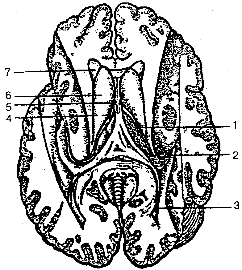
Rice. 115. Lateral ventricles of the cerebrum (opened),
horizontal section:
1 - central part of the lateral ventricle; 2 - lower horn; 3 - posterior horn; 4 - interventricular foramen; 5 - transparent partition; 6 - head of the caudate nucleus; 7- front horn
The ventricular cavity is different in shape, since it is formed by sections of all lobes of the hemispheres (except for the insular one). The central part of the ventricle lies in the parietal lobe. From the central part, processes - horns - diverge into all lobes of the brain: anterior (frontal) - into the frontal lobe; lower (temporal) - into the temporal; posterior (occipital) - into the occipital lobe.
The lateral ventricles are closed on all sides, with the exception of the interventricular foramen, through which they are connected to the third ventricle, and with its help - to each other.
Third ventricle - unpaired slit-shaped cavity, located in the diencephalon. The cavity of this ventricle is limited by six walls: two lateral, upper, lower, middle and posterior. The lateral walls are medial surfaces thalamus. The upper wall is formed by the vascular base (soft choroid). The lower wall, or bottom, of the third ventricle is the hypothalamus. The anterior wall is formed by the lamina terminalis, the pillars of the fornix and the anterior commissure. Through the interventricular foramen, the cavity of the third ventricle is connected to the lateral ventricles. The posterior wall is the epithalamic commissure, under which is the opening of the cerebral aqueduct.
Fourth ventricle is a derivative of the cavity of the rhombencephalon (Fig. 116). The shape of the cavity of the IV ventricle resembles a tent, the bottom of which has the shape of a diamond (diamond-shaped fossa), formed by the posterior surfaces of the medulla oblongata and the pons, as well as the cerebellum and the isthmus of the rhombencephalon. The cavity of the fourth ventricle is connected to the subarachnoid space by three openings - an unpaired middle and paired lateral.
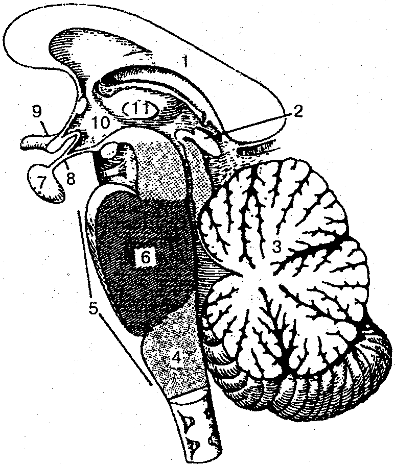
Rice. 116. Rhombencephalon (sagittal section):
1 - corpus callosum; 2- pineal body (epiphysis); 3 - cerebellum; 4 - medulla; 5 - rhomboid brain; 6- bridge; 7- pituitary gland; 8- funnel; 9- visual chiasm; 10 - hypothalamus; 11 - intergalamic fusion
The membranes of the brain. The brain is surrounded by three membranes, which are a continuation of the membranes of the spinal cord (Fig. 117).
Dura shell the brain is at the same time the periosteum of the inner surface of the bones of the skull, with which it is loosely connected. At the base of the skull, the membrane gives off processes that penetrate into the cracks and openings of the skull. On the inner surface of the dura mater, several processes are distinguished that penetrate into the deep crevices of the brain and separate its sections. The largest process of the dura mater of the brain between the hemispheres is falx cerebri. The posterior part of the falx fuses with another process of the dura mater - the tentorium cerebellum, which separates the occipital lobes of the hemispheres from the cerebellum. The continuation of the falx cerebri is falx cerebellum, which penetrates between the cerebellar hemispheres. Another process surrounds the sella turcica, forms its diaphragm and protects the pituitary gland from the pressure of the brain mass.

Rice. 117. Meninges of the brain:
1 - granulation of the arachnoid membrane; 2- emissary vein; 3- vein of cancellous bone; 4 - dura mater; 5 - crossbars of the arachnoid membrane; 6 - subarachnoid space; 7-vascular membrane; 8 - arachnoid; 9 - falx cerebri; 10 - superior sagittal sinus; 11 - cerebral cortex
In the corresponding areas of the dura mater of the brain there are sinuses (sinuses), formed by splitting the dura mater; Venous blood flows through these sinuses. The following sinuses are distinguished: 1) superior sagittal; 2) lower sagittal; 3) straight; 4) transverse; 5) occipital; b) sigmoid; 7) cavernous; 8) sphenoparietal; 9) upper and lower rocky.
Arachnoid membrane of the brain located inside the dura mater and separated from it by the subdural space. The arachnoid membrane in the form of bridges is transferred from one part to another. The arachnoid is separated from the soft membrane by the subarachnoid (subarachnoid) space, which contains cerebrospinal fluid. Above the wide and deep grooves, the arachnoid membrane forms subarachnoid cisterns. Of these, the largest are: 1) the cerebellocerebral cistern; 2) cistern of the lateral fossa cerebri; 3) crossover tank; 4) interpeduncular cistern.
The subarachnoid space of the brain connects with the subarachnoid space of the spinal cord at the level of the foramen magnum. Near the sinuses of the dura mater of the brain, the arachnoid membrane forms peculiar outgrowths - granulations of the arachnoid membrane. These outgrowths enter the sinuses of the dura mater.
Soft (vascular) membrane - the innermost layer of the brain. It fits tightly to the surface of the brain, extends into all the cracks and grooves. It consists of loose connective tissue, in the thickness of which there are blood vessels that provide nutrition to the brain. In some places, the choroid forms choroid plexuses that produce cerebrospinal fluid.
Cerebrospinal fluid - a liquid biological environment of the body that circulates in the ventricles of the brain, the cerebrospinal fluid ducts, and the subarachnoid space of the brain and spinal cord. It performs a protective-trophic function in the central nervous system, participates in brain metabolism, etc.
The total volume of cerebrospinal fluid in an adult is on average 140 ml. It is updated approximately 4-8 times a day and depends on nutrition, water regime, physical activity and etc. Chemical composition Cerebrospinal fluid is similar to the composition of blood serum; it contains organic and inorganic substances that take part in brain metabolism. With various pathological processes in the central nervous system, changes in fluid pressure, its properties and composition are possible, which reflect a particular disease.
| |