Furrows and convolutions of the upper lateral surface. Large hemispheres. Major lobes, gyri and sulci of the cerebral hemispheres. Elements of the functional organization of the hemispheres
The last functional part of the brain covers the anterior half hemispheres. Here are the centers that control our actions.
From here, nerve pathways lead to the muscles of the limbs, face, lips, eyes, and tongue. Impulses sent out from "motor" centers in the cortex allow us to move, speak and control facial expressions - we blink, smile, frown and pout.
Although the cerebral hemispheres seem exactly the same, they nevertheless perform different functions. They are connected to each other by a strip of nerve tissue called the corpus callosum. If it is removed or damaged, both hemispheres act relatively independently of each other, generating their own thoughts and emotions.
Left hemisphere controls movements right side body. Here are the most important centers of speech, language, mathematical abilities and logical thinking. Right hemisphere responsible for left side and is the focus of visual perception, musical talent and abstract thinking. In this case, there is a continuous exchange of impulses between the hemispheres, and even if the corpus callosum is cut, both halves of the brain still operate more or less smoothly.
Bark big brain covers the surface of the hemispheres and forms a large number of furrows of various depths and lengths (lat. sulci cerebri). Between the furrows are located various sizes of the gyrus of the brain (Latin gyri cerebri)
In each hemisphere, the following surfaces are distinguished:
convex upper lateral surface (lat. facies superolateralis), adjacent to the inner surface of the bones of the cranial vault
lower surface (lat. facies inferior), the anterior and middle sections of which are located on the inner surface of the base of the skull, in the region of the anterior and middle cranial fossae, and the posterior ones - on the cerebellum
medial surface (lat. facies medialis), directed to the longitudinal fissure of the brain
These three surfaces of each hemisphere, passing one into another, form three edges. The upper edge (Latin margo superior) separates the upper lateral and medial surfaces. The inferolateral edge (Latin margo inferolateralis) separates the upper lateral surface from the lower one. Inferomedial edge (Latin margo inferomedialis) is located between the lower and medial surfaces
In each hemisphere, the most protruding places are distinguished: in front - the frontal pole (Latin polus frontalis), behind - the occipital (Latin polus occipitalis), and on the side - temporal (Latin polus temporalis)
The hemisphere is divided into five lobes. Four of them are adjacent to the corresponding bones of the cranial vault:
frontal lobe (lat. lobus frontalis)
parietal lobe (Latin lobus parietalis)
occipital lobe (Latin lobus occipitalis)
temporal lobe (Latin lobus temporalis)
Fifth - insular lobe (lat. Lobus insularis) (islet) (lat. insula) - is laid in the depths of the lateral fossa of the brain (lat. Fossa lateralis cerebri), which separates the frontal lobe from the temporal lobe
Furrows and convolutions of the upper lateral surface
frontal lobe- marked in pink
Parietal lobe - marked in green
Temporal lobe - marked in yellow
Occipital lobe - marked in blue
frontal lobe
The frontal lobe is separated from the parietal by a deep central sulcus (lat. sulcus centralis). It starts on medial surface hemisphere, passes to its upper lateral surface, goes along it a little obliquely, from back to front, and usually does not reach lateral furrow brain
Approximately parallel to the central sulcus is the precentral sulcus (Latin sulcus precentralis), which does not reach the upper edge of the hemisphere. The precentral sulcus borders the precentral gyrus (Latin gyrus precentralis) in front.
The upper and lower frontal sulci (lat. sulci frontales superior et inferior) are directed forward from the precentral sulcus. They divide the frontal lobe into:
superior frontal gyrus (lat. gyrus frontalis superior), which is located above the superior frontal sulcus and passes to the medial surface of the hemisphere
the middle frontal gyrus (Latin gyrus frontalis medius), which is limited by the upper and lower frontal sulci. The orbital (anterior) segment of this gyrus passes to the lower surface of the frontal lobe
the lower frontal gyrus (lat. gyrus frontalis inferior), which lies between the lower frontal sulcus and the lateral sulcus of the brain and the branches of the lateral sulcus is divided into a number of parts
The lateral sulcus (lat. Sulcus lateralis) is one of the deepest sulci of the brain. It separates the temporal lobe from the frontal and parietal. The lateral groove lies on the upper lateral surface of each hemisphere and goes from top to bottom and anteriorly. In the depths of this groove there is a recess - the lateral fossa of the brain (lat. Fossa lateralis cerebri), the bottom of which is the outer surface of the island
Small furrows, called branches, depart upward from the lateral furrow. The most constant of them are the ascending (lat. ramus ascendens) and the anterior (lat. ramus anterior) branches. The upper-posterior part of the furrow is called the posterior branch (lat. ramus posterior)
The inferior frontal gyrus, within which the ascending and anterior branches pass, is divided by them into three parts:
back - tire part (lat. pars opercularis), bounded in front by an ascending branch
middle - triangular part (lat. pars triangularis), lying between the ascending and anterior branches
anterior - orbital part (lat. pars orbitalis), located between the anterior branch and the inferolateral edge of the frontal lobe
parietal lobe
It lies behind the central sulcus, which separates it from the frontal sulcus. It is delimited from the temporal by the lateral sulcus of the brain, from the occipital by a part of the parietal-occipital sulcus (lat. sulcus parietooccipitalis)
Parallel to the precentral gyrus, the postcentral gyrus runs (lat. Gyrus postcentralis). Posteriorly from it, almost parallel to the longitudinal fissure of the large brain, there is an intraparietal sulcus (lat. sulcus intraparietalis), dividing the posterior superior sections of the parietal sections of the parietal lobe into two gyrus: upper (lat. lobulus parietalis superior) and lower (lat. lobulus parietalis inferior) parietal slices. In the lower parietal lobule, two relatively small convolutions are distinguished: supramarginal (lat. gyrus supramarginalis), lying anteriorly and closing the posterior parts of the lateral sulcus, and located posterior to the previous angular (lat. gyrus angularis), which closes the superior temporal sulcus
Between the ascending and posterior branches of the lateral sulcus of the brain is a section of the cortex, referred to as the fronto-parietal tire (Latin operculum frontoparietalis). It includes the posterior part of the inferior frontal gyrus, the lower sections of the precentral and postcentral gyri, as well as the lower section of the anterior part of the parietal lobe.
Occipital lobe
On the upper lateral surface, it has no boundaries separating it from the parietal and temporal lobes, with the exception of the upper part of the parietal-occipital sulcus, which is located on the medial surface of the hemisphere and separates the occipital lobe from the parietal
The largest of the furrows is the transverse occipital sulcus (Latin sulcus occipitalis transversus). Sometimes it is a continuation of the posterior intraparietal sulcus and in the posterior section passes into an unstable semilunar sulcus (Latin sulcus lunatus)
temporal lobe
Has the most pronounced boundaries. It distinguishes between a convex lateral surface and a concave lower one. The obtuse pole of the temporal lobe is turned forward and somewhat downward. The lateral sulcus of the brain sharply delimits the temporal lobe from the frontal lobe.
Two grooves located on the upper lateral surface: the upper (lat. sulcus temporalis superior) and the lower (lat. sulcus temporalis inferior) temporal grooves, following almost parallel to the lateral groove of the brain, divide the lobe into three temporal gyrus: superior, middle and inferior (lat. gyri temporales superior, medius et inferior)
Those parts of the temporal lobe that are directed towards the lateral sulcus of the brain are indented with short transverse temporal sulci (Latin sulci temporales transversi). Between these furrows lie 2-3 short transverse temporal gyri associated with the gyri of the temporal lobe (Latin gyri temporales transversi) and the insula
Islet share (islet)
It lies at the bottom of the lateral fossa of the brain (Latin fossa lateralis cerebri).
It is a three-sided pyramid, turned by its top - the pole of the island - anteriorly and outwards, towards the lateral groove. From the periphery, the islet is surrounded by the frontal, parietal and temporal lobes, which are involved in the formation of the walls of the lateral sulcus of the brain.
The base of the island is surrounded on three sides by a circular furrow of the island (lat. sulcus circularis insulae).
Its surface is cut by a deep central groove of the island (Latin sulcus centralis insulae). This furrow divides the islet into anterior and posterior parts.
On the surface, a large number of small convolutions of the island (Latin gyri insulae) are distinguished. The large anterior part consists of several short convolutions of the insula (lat. gyri breves insulae), the posterior part consists of one long convolution (lat. gyrus longus insulae)
hemispheres (Figure 10)
Furrows of the first order divide each hemisphere into lobes. Lateral (Sylvian) furrow separates the temporal and parietal lobes. Central (Roland's) furrow separates the frontal and parietal lobes. Occipito-parietal sulcus separates the occipital and parietal lobes. Furrows of the second order divide each lobe of the hemisphere into convolutions.
frontal lobe. It has precentral, superior, and inferior sulci, and, accordingly, the anterior central, superior, middle, and inferior frontal gyri. In addition, there are branches of the lateral sulcus - anterior and ascending, which divide the inferior frontal gyrus into orbital , triangular And tire parts .
parietal lobe. It has postcentral, interparietal grooves and, accordingly, the posterior central, superior and inferior parietal gyrus. The part of the inferior parietal gyrus that surrounds the lateral sulcus is called supramarginal gyrus , the other part goes around the upper temporal, is called supraangular gyrus .
temporal lobe. It has superior and inferior sulci and, correspondingly, superior, middle, and inferior temporal gyrus.
Occipital lobe. The main furrow is transverse (spur).
Island. It is located under the lateral groove, has the shape of a triangle (Figure 13).
Furrows and convolutions of the mediobasal surface of the hemisphere(pictures 11, 12)
Figure 11. Medial surface of the right hemisphere of the brain: 1 - paracentral lobule; 2 - prewedge; 3 - occipito-parietal sulcus; 4 - wedge; 5 - gyrus of the hippocampus (parahippocampal); 6 - hook; 7 - cingulate gyrus; 8 - transverse (spur furrow); 9 - corpus callosum
Olfactory brain (Figures 11, 12). Consists of peripheral and central sections. Peripheral - olfactory bulbs, tracts, triangles and anterior perforated substance. Central part - gyrus of Ammonovarog (hippocampus, sea horse), dentate, vaulted gyrus and hooks. The olfactory brain is part of the limbic system.

Figure 12. The lower surface of the brain: 1 - mastoid bodies; 2 - olfactory bulbs; 3 - olfactory tracts; 4 - olfactory triangles; 5 - anterior perforated space; 6 - gyrus of the hippocampus; 7 - hook; 8 - occipital-temporal lateral (piri-shaped) gyrus; 9 - occipital-temporal medial (lingual) gyrus
Lateral ventricles of the brain(Figure 13). Found in every hemisphere. First on the left, second on the right. Their parts form the anterior, inferior and posterior horns.
Basal nuclei of the hemispheres(Figure 13). The accumulation of gray matter in its thickness or "subcortex". Form the striatal system ( striatum ) and a system of pale balls ( pallidum ).

In addition to these nuclei, the basal nuclei include fence And almond-shaped nuclei . Each of these nuclei also has its own specific functions.
Tailed nuclei. Regulate the transition from one type of movement to another.
Shell. Pair education. Organizes motor activity, participates in the organization of eating behavior and in its integration with the functions of respiration and salivation.
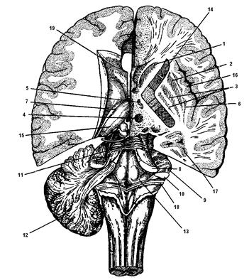
Figure 13. Hemispheres of the brain on different levels horizontal section (on the right - below the level of the bottom of the lateral ventricle, on the left - above the bottom of the lateral ventricle): 1 - caudate nucleus; 2 - shell; 3 - pale balls; 4 - red nuclei; 5 - subthalamic body of Lewis; 6 - fence; 7 - almond-shaped nucleus; 8 - upper legs of the cerebellum; 9 - middle legs of the cerebellum; 10 - lower legs of the cerebellum; 11 - upper brain sail; 12 - cerebellum; 13 - diamond-shaped fossa; 14 - inner capsule; 15 - thalamus; 16 - islet bark; 17 - lower horn; 18 - brain strips; 19 - front horn
pale balls. They regulate the launch or activation of the orienting reaction, limb movements and eating behavior (chewing, swallowing).
Fence. Pair education. Participates in excitatory reactions to somatic, auditory, visual stimuli (orienting reactions, turning the head, chewing, swallowing, vomiting movements).
Almond nucleus. Pair education. Located deep in the temporal lobe. Participates in defensive, vegetative, motor and emotional reactions. The striopallidar system is part of the extrapyramidal system.
(page 7 of 31)Anterior to the optic chiasm is the terminal plate, lamina terminalis, which is located in the frontal plane and is a continuation of the ventral end of the corpus callosum. It ends at the base of the knee of the corpus callosum.
The ventricular surface of the hypothalamus has two depressions along the median plane. The anterior is located between the optic chiasm and the terminal plate - supraoptic recess, recessus supraopticus. The second recess corresponds to the funnel - the recess of the funnel, recessus fundibuli.
telencephalon, telencephalon, is a derivative of the anterior cerebral bladder and is represented by two cerebral hemispheres, hemispheria cerebrales. In each hemisphere there are:
1) raincoat, pallium, formed from the dorsal wall of the brain bladder;
2) basal nuclei, nuclei basales developing from its ventral region.
Inside each hemisphere there is a cavity - the lateral ventricle, ventriculus lateralis.
outer layer raincoat is the cerebral cortex, cortex cerebri, under which the white matter is located, which makes up the largest part of the hemisphere. Functionally telencephalon contains higher integration centers responsible for conscious sensitivity, voluntary movements, mental ability and memory.
Basal nuclei, nuclei basales, are a group of nuclei located at the base of each hemisphere. The entire group of basal nuclei makes up a mass of gray matter, which is generally ovoid in shape.
The basal ganglia include: caudate nucleus, nucleus caudatus; lenticular nucleus, nucleus lentiformis; fence, claustrum, and the amygdala, corpus amygdaloideum.
The caudate nucleus has the shape of a comma located in the sagittal plane, with a longitudinally oriented long axis. It is located laterally and above the thalamus. The anterior end of the caudate nucleus is the head, caput, has a thickening. Gradually decreasing in volume, the head of the nucleus continues into the body, corpus, the free surface of which protrudes into the cavity of the lateral ventricle. The body of the caudate nucleus, gradually thinning and curving down, continues into the tail, cauda. The caudate nucleus, with its bend, encloses the fibers white matter, partially continuing from the legs of the brain. The size of the caudate nucleus in the sagittal direction reaches 6–7 cm. The greatest width in the region of the head is 20 mm, and in the region of the tail it is approximately 3 mm.
Lateral to the caudate nucleus and the thalamus is a well-defined strip of white matter - the internal capsule, capsula interna, the width of which is 5 - 7 mm. The internal capsule separates the caudate nucleus from the lenticular nucleus. lenticular nucleus, nucleus lentiformis, is surrounded on all sides by white matter and has a wedge-shaped shape in all planes.
It has two parts - lateral and medial. The lateral part, which is larger in size, is called the shell, putamen; the medial part is a pale ball, Globus pallidus.
Shell, putamen, like the caudate nucleus, has a cepo-pink color. pale ball, Globus pallidus, on a fresh preparation differs in yellowish color. thin layer of white matter, the cerebral lamina lamina medullaris, separates the shell from the pale ball.
Fence, claustrum, is located lateral to the shell and is separated from it by a layer of white matter, representing the outer capsule, capsula externa. Even more lateral is a strip of white matter - the outermost capsule, capsula extrema separating the fence from the bark of the island. The fence on the horizontal section of the hemisphere looks like a thin strip of gray matter (average 1 - 2 mm). Its outer surface has jagged contours corresponding to the gyri of the insular cortex. In the direction upwards and downwards, the fence becomes thinner and approaches the amygdala. In a three-dimensional image, it looks like a disk located in the sagittal plane.
amygdala, corpus amygdaloideum, in shape and size (about 10 mm) resembles an almond pit. It is located in the thickness of the white matter of the pole of the temporal lobe. The amygdala with its upper surface acts as an elevation in the anterior section of the lower horn of the lateral ventricle. Thin plates of white matter, it is divided into a number of secondary nuclei.
cerebral cortex It is a layer of gray matter, the thickness of which varies in different departments and averages 2-3 mm. In an adult, due to the final formation of furrows, the area of \u200b\u200bthe cerebral hemispheres averages 1550 cm 2. The surface of the bark has a complex relief, characterized by numerous furrows, Sulci Cerebri, and the elevations located between them - convolutions, gyri cerebri. The convolutions differ from each other in shape and size. There is a pronounced individual variability in the relief of the cerebral cortex, however, the sulci and gyrus of the same name in various people fundamentally similar and localized in certain places.
The founder of the study of the cellular composition of the cerebral cortex, the features of the structure and distribution of nerve cells (cytoarchitectonics of the cortex) is Professor of Kyiv University V. A. Bets. Later, in the cerebral cortex, Korbinian Brodman identified 52 fields, designating each of them with a certain number. The same numbering of fields is preserved in the cytoarchitectonic map compiled by the Institute of the Brain of Russia, but on it a number of fields are subdivided into zones designated by letters of the Latin alphabet.
In each hemisphere of the large brain, the upper lateral, medial and lower surfaces are distinguished. The upper lateral surface of the hemispheres is the most extensive, has a convex shape, faces upward and laterally. It borders on the medial surface with a clearly defined edge. The flat medial surface faces the longitudinal fissure of the brain, , in the middle part is connected by the corpus callosum with the same surface of the other hemisphere. The lower surface is flattened in the anterior part, and concave in the posterior part. Three major sulci divide each hemisphere into four lobes, Lobi Cerebri.
1. Lateral (lateral) furrow, sulcus lateralis, begins on the lower surface of the hemisphere in the form of a lateral (Sylvian) fossa of the brain, fossa lateralis cerebri (Sylvii), goes along the lateral side up and back. It is the anterior superior border of the temporal lobe, lobus temporalis, and separates the frontal and parietal lobes from the temporal.
2. Central sulcus, sulcus centralis (Rolandi), runs along the upper lateral surface of the hemisphere, starting from its upper edge. Usually it passes to its medial side and bottom slightly does not reach the lateral furrow. The central sulcus divides the upper part of the hemisphere into anterior section, including the frontal lobe, lobus frontalis, and posterior, including the parietal lobe, lobus parietalis, and the occipital lobe, lobus occipitalis. characteristic feature the central sulcus is its continuity throughout.
3. Parieto-occipital sulcus, sulcus parietooccipitalis, is located in the back of the brain on the medial surface of the hemisphere, continuing slightly to the upper lateral surface. This groove is the boundary between the parietal and occipital lobes.
In addition to the main four lobes, there is also an island, insula (Reilli), which is also called the insular lobe, lobus insularis. It lies in the depths of the lateral furrow and is visible only when the convolutions that limit this furrow are pulled apart.
Furrows and convolutions of the upper lateral surface of the cerebral hemispheres(Fig. 16). frontal lobe, lobus frontalis. On the upper lateral surface anterior to the central sulcus are the superior precentral sulcus and the inferior precentral sulcus. More often they merge into one precentral sulcus, sulcus precentralis. From this furrow originate, heading forward, two frontal furrows: upper, sulcus frontalis superior, and lower, sulcus frontalis inferior.
These furrows describe the surface of the frontal lobe is divided into convolutions. Anterior to the central sulcus is the precentral gyrus, gyrus precentralis. Three frontal gyri are distinguished in the rest of the area:
- superior frontal gyrus gyrus frontalis superior, which is above sulcus frontalis superior along the upper edge of the hemisphere;
- middle frontal gyrus gyrus frontalis medius, which lies between the upper and lower frontal furrows;
- inferior frontal gyrus gyrus frontalis inferior, is between sulcus frontalis inferior And sulcus lateralis.
parietal lobe, lobus parietalis. On the upper lateral surface, the postcentral sulcus runs parallel to the central sulcus, sulcus postcentralis. From it begins in the sagittal direction a long intraparietal sulcus, sulcus intraparietalis. These two furrows divide the surface of the parietal lobe into three sections. The postcentral gyrus is located between the central and postcentral sulci. gyrus postcentralis. Above, it continues to the medial surface of the hemisphere. The area of the cortex located above sulcus intraparietalis, is called the superior parietal lobule, lobulus parietalis superior. The underlying area is the lower parietal lobule, lobulus parietalis inferior. It contains two very important gyrus: supramarginal, gyrus supramarginalis, surrounding the rear end sulcus lateralis, and angular, gyrus angularis, closing the end sulcus temporalis superior.
Rice. 16. The relief of the upper lateral surface of the cerebral hemispheres:
1 – sulcus precentralis; 2 - sulcus centralis; 3 - sulcus postcentralis; 4 - sulcus intraparietalis; 5 - sulcus parietooccipitalis; 6 - gyri occipitales superiores; 7 - sulcus occipitalis transversus; 8 - gyri occipitales laterales; 9 – sulcus temporalis inferior; 10 - sulcus temporalis superior; 11 - fossa lateralis cerebri (Sylvii); 12 - sulcus frontalis inferior; 13 - sulcus frontalis superior
Occipital lobe, lobus occipitalis, is the smallest of all shares. On the upper lateral surface, its furrows vary greatly. Here, a semilunar furrow is distinguished, sulcus lunatus, transverse occipital sulcus, sulcus occipitalis transversus, as well as pre-occipital notch, incisura preoccipitalis, which delimits this lobe from the temporal lobe from below. Conventionally, on the upper lateral surface, it is possible to distinguish the superior and lateral occipital gyri, gyri occipitales superiores and laterales.
temporal lobe, lobus temporalis. On the upper lateral surface in the anteroposterior direction passes the superior temporal groove, sulcus temporalis superior, which, with its posterior end, extends into the region of the parietal lobe. inferior temporal sulcus, sulcus temporalis inferior, is best seen from the bottom surface.
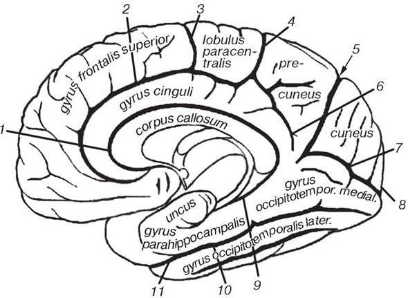
Rice. 17. The relief of the medial surface of the cerebral hemispheres:
1 – sulcus corporis callosi; 2 - sulcus cinguli; 3 - sulcus paracentralis; 4 - ramus marginalis; 5 - sulcus parietooccipitalis; 6 - sulcus subparietalis; 7 - sulcus calcarinus; 8 - polus occipitalis; 9 - sulcus hippocampalis; 10 - sulcus collateralis; 11 - sulcus rhinalis
On the upper lateral surface of the temporal lobe are the superior temporal gyrus, gyrus temporalis superior, and the middle temporal gyrus, gyrus temporalis medius. They are separated from each other by the superior temporal groove. Along the lower edge of the hemisphere runs the inferior temporal gyrus, gyrus temporalis inferior bounded by the inferior temporal sulcus.
At the frontal, temporal and occipital lobe and distinguish the most protruding poles of the same name ( polus frontalis, polus temporalis, polus occipitalis).
Island, insula (Reilii), clearly visible only when diluting the frontal / parietal and temporal lobes, limiting the furrow sulcus lateralis (Sylvii) at the bottom of which it is located. The islet bears some resemblance to a cone, the base of which is surrounded by a deep circular furrow of the islet, sulcus circularis insulae. Its surface is divided by the central sulcus of the insula, sulcus centralis insulae, on the anterior and posterior lobes. The posterior lobe usually consists of only one long gyrus of the insula, gyrus longus insulae, anterior contains several short insular convolutions, gyri breves insulae.
Furrows and convolutions of the medial surface of the cerebral hemispheres(Fig. 17). All lobes of the cerebral hemispheres are continued on the medial surface. The main sulcus here is the sulcus of the corpus callosum, sulcus corporis callosi, which surrounds the corpus callosum on its convex side. Approximately halfway between sulcus corporis callosi and the upper edge of the hemisphere is the cingulate groove, Sulcus cinguli. It turns to the upper edge of the hemisphere with its posterior end - the marginal branch, ramus marginalis, and slightly extends onto the dorsolateral surface, posterior to the central sulcus. In front of the marginal branch, approximately above the middle of the corpus callosum, the cingulate gyrus gives up the paracentral sulcus, sulcus paracentralis. The immediate continuation of the cingulate sulcus is the subtopic sulcus, sulcus subparietalis. Below the posterior end of the corpus callosum, two grooves begin with a common trunk, diverging towards the edge of the hemisphere: the already described parietal-occipital, sulcus parietooccipitalis, and spur furrow, sulcus calcarinus. Near the occipital pole, on the lower surface of the hemisphere, a collateral groove begins, sulcus collateralis heading forward. Its continuation in the anterior part of the temporal lobe is the olfactory groove, sulcus rhinalis. Lateral to the collateral sulcus is the occipitotemporal sulcus, sulcus occipitotemporalis.
The part of the medial surface that lies above the cingulate gyrus belongs to the frontal lobe - this is the superior frontal gyrus that extends here. Behind, it reaches the level of the projection of the upper end of the central sulcus. Within the parietal lobe is the paracentral lobule, lobulus paracentralis, which at the bottom reaches the subparietal sulcus, sulcus subparietalis. The paracentral lobule connects the parietal lobe with the frontal lobe on the medial surface (more precisely, gyrus postcentralis With gyrus precentralis). Between pars marginalis sulci cinguli- in front, sulcus parietooccipitalis- behind and sulcus subparietalis- below lies the prewedge, precuneus. Between sulcus parietooccipitalis And sulcus calcarinus(already in the occipital lobe) there is a wedge, cuneus. On the medial surface of the same lobe is the lingual gyrus, gyrus lingualis lying between sulcus calcarinus And sulcus collateralis. Below the latter is the medial occipitotemporal gyrus, gyrus occipitotemporalis medialis.
Within the temporal lobe on the medial surface of the hemispheres directly under the legs of the brain is the parahippocampal gyrus, gyrus parahippocampalis, which ends with a hook in front, uncus. From the legs of the brain, the parahippocampal gyrus and the hook are separated by the hippocampal groove, sulcus hippocampalis. Below the parahippocampal gyrus lies the lateral occipitotemporal gyrus, gyrus occipitotemporalis lateralis. The named convolutions are separated behind by a collateral groove, sulcus collateralis, in front - olfactory groove, sulcus rhinalis.
The inferior temporal gyrus runs along the lowest edge of the medial surface of the temporal lobe, gyrus temporalis inferior, which is separated by the occipitotemporal sulcus, sulcus occipitotemporalis, from the lateral occipitotemporal gyrus.
The convolutions, annularly bordering the corpus callosum and the legs of the brain, extending from the frontal lobe to the temporal lobe, as a whole make up the limbic lobe, lobus limbicus. It consists of two parts: the cingulate gyrus, gyrus cinguli, and the parahippocampal gyrus, gyrus parahippocampalis connected to each other by an isthmus, isthmus gyri cinguli, which begins behind the roller of the corpus callosum. The cingulate gyrus lies between the corpus callosum sulcus, on the one hand, and the cingulate sulcus and subparietal sulcus, on the other. The parahippocampal gyrus, as already noted, is limited from above by the hippocampal sulcus, sulcus hippocampalis, below - the anterior end of the collateral and olfactory furrows.
The relief of the lower surface of the hemispheres(Fig. 18). On the lower (basal) surface of the frontal lobe is the olfactory groove, sulcus olfactorius running parallel to the longitudinal fissure of the brain. The olfactory tract passes through the furrow. More lateral to the olfactory sulcus of the frontal lobe are the orbital sulci, sulci orbitales. Between these furrows there are convolutions of variable shape: a direct gyrus, gyrus rectus, which is limited sulcus olfactorius And fissura longitudinalis cerebri, and orbital gyri, gyri orbitales lying laterally from the olfactory sulcus.
Within the temporal and occipital lobes there is no clear boundary between the medial and inferior surfaces. They gradually pass into each other. In this regard, the furrows and convolutions located on the medial surface of the hemispheres in the lower parts of the occipital and temporal lobes are also visible on the lower surface of the hemispheres. In particular, within occipital lobe is the medial occipitotemporal gyrus. Within the temporal lobe lie the parahippocampal, lateral occipitotemporal, and inferior temporal gyrus. The sequence of location of these convolutions was considered in the lateral direction. The furrows separating these convolutions were named earlier.
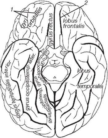
Rice. 18. The relief of the lower surface of the cerebral hemispheres:
1- sulci orbitales; 2 - sulcus olfactorius
The above description of the furrows and convolutions of the cerebral cortex is schematic, since individual variants of their architectonics are quite common.
Projection and associative centers of the cerebral cortex. The physiological permissibility of surgical interventions on the brain in most cases is determined by the localization of nerve centers in the cerebral cortex. Projection centers are areas of the cerebral cortex, which are the cortical representation of the analyzer and have a direct morphofunctional connection through afferent or efferent nerve pathways with the neurons of the subcortical centers. Associative centers are areas of the cerebral cortex that do not have a direct connection with subcortical formations, but are connected by a temporary two-way connection with projection centers. Associative centers play a paramount role in the implementation of the highest nervous activity. Within the framework of this publication, we will not dwell on a detailed description of each of the centers, but will only give their location in the cerebral cortex.
The projection center of general sensitivity (tactile, pain, temperature and conscious proprioceptive) is also called the skin analyzer of general sensitivity. It is localized in the cortex of the postcentral gyrus.
The projection center of motor functions (kinesthetic center), or motor analyzer, is located in the motor area of the cortex, which includes the precentral gyrus and the paracentral lobule.
The projection center of hearing, or the core of the auditory analyzer, is located in the middle third of the superior temporal gyrus, mainly on the surface of the gyrus facing the insula.
The projection center of vision, or the core of the visual analyzer, is localized on the medial surface of the occipital lobe, along the edges of the spur groove.
The projection center of smell, or the core of the olfactory analyzer, is located on the medial surface of the temporal lobe, in the cortex of the parahippocampal gyrus and in the hook (limbic region).
The projection center of taste, or the core of the taste analyzer, is located in the same place as the projection center of smell, that is, in the limbic region of the brain.
Projection center sensitivity from internal organs, or analyzer of visceroception, is located in the lower third of the postcentral and precentral gyri.
The projection center of vestibular functions, the vestibular analyzer, undoubtedly has its representation in the cerebral cortex, but information about its localization is ambiguous. It is generally accepted that the projection center of vestibular functions is located on the lateral surface of the temporal lobe, in the region of the middle and inferior temporal gyri.
The associative center of stereognosia, or the core of the skin analyzer of object recognition by touch, is located in the superior parietal lobe.
The associative center of the body scheme is located in parietal lobe, in the region of the intraparietal sulcus.
The associative center of praxia, or the analyzer of purposeful habitual movements, is located in the lower parietal lobule, in the cortex of the supramarginal gyrus, in right-handers - in the left hemisphere of the brain, in left-handers - in the right. The associative center of vision, or the analyzer of visual memory, is located on the upper lateral surface of the occipital lobe, for right-handers - in the left hemisphere, for left-handers - in the right.
The associative center of hearing, or the acoustic center of speech, is also called the Wernicke center (by the name of the German neurologist and psychiatrist Karl Wernicke, who first described in 1874 the symptoms of damage to the posterior third of the superior temporal gyrus, within which this center is located on the dominant hemisphere).
The associative motor center of speech (speech motor), or the center of speech articulation, is called Broca's center (by the name of Paul Broca, a French anatomist and surgeon, who in 1861 for the first time demonstrated at a meeting of the Paris Anthropological Society the brain of a patient who had motor aphasia during his lifetime , with a lesion in the region of the posterior third of the inferior frontal gyrus). The center is located on the dominant hemisphere.
Presented here introductory fragment books.
Only part of the text is open for free reading (restriction of the copyright holder). If you liked the book full text can be obtained from our partner's website.